. 1999 Oct;67(10):5282-91.
doi: 10.1128/IAI.67.10.5282-5291.1999.

Cryptosporidium parvum apical complex glycoprotein CSL contains a sporozoite ligand for intestinal epithelial cells

Affiliations

Cryptosporidium parvum apical complex glycoprotein CSL contains a sporozoite ligand for intestinal epithelial cells

R C Langer et al. Infect Immun. 1999 Oct.

Abstract

Cryptosporidiosis, caused by the apicomplexan parasite Cryptosporidium parvum, has become a well-recognized diarrheal disease of humans and other mammals throughout the world. No approved parasite-specific drugs, vaccines, or immunotherapies for control of the disease are currently available, although passive immunization with C. parvum-specific antibodies has some efficacy in immunocompromised and neonatal hosts. We previously reported that CSL, an approximately 1,300-kDa conserved apical glycoprotein of C. parvum sporozoites and merozoites, is the antigenic species mechanistically bound by neutralizing monoclonal antibody 3E2 which elicits the circumsporozoite precipitate (CSP)-like reaction and passively protects against C. parvum infection in vivo. These findings indicated that CSL has a functional role in sporozoite infectivity. Here we report that CSL has properties consistent with being a sporozoite ligand for intestinal epithelial cells. For these studies, native CSL was isolated from whole sporozoites by isoelectric focusing (IEF) following observations that the approximately 1,300-kDa region containing CSL as seen by sodium dodecyl sulfate-polyacrylamide gel electrophoresis was comprised of approximately 15 molecular species (pI 3 to 10) when examined by two-dimensional (2-D) electrophoresis and silver staining. A subset of six approximately 1,300-kDa species (pI 4.0 to 6.5) was specifically recognized by 3E2 in 2-D Western immunoblots of IEF-isolated CSL. Isolated native CSL bound specifically and with high affinity to permissive human intestinal epithelial Caco-2 cells in a dose-dependent, saturable, and self-displaceable manner. Further, CSL specifically bound to the surface of live Caco-2 cells inhibited sporozoite attachment and invasion. In addition, sporozoites having released CSL after incubation with 3E2 and occurrence of the CSP-like reaction did not attach to and invade Caco-2 cells. These findings indicate that CSL contains a sporozoite ligand which facilitates attachment to and invasion of Caco-2 cells and, further, that ligand function may be disrupted by CSL-reactive monoclonal antibody. We conclude that CSL is a rational target for passive or active immunization against cryptosporidiosis.


Figures

FIG. 1
FIG. 1
(A) Silver-stained 2 to 12% gradient SDS-PAGE gel of solubilized oocysts before (lane 1, 105) and after (lane 2, 1.4 μg) IEF isolation of CSL (arrow). Lane 3 was loaded with sample buffer to identify silver-stain artifacts. (B) Western blot recognition of IEF-isolated CSL (lane 2, 0.5 μg) (arrow) and an ∼1,300-kDa comigrating antigen in solubilized oocysts (lane 1, 7 × 10⁶) by MAb 3E2. Lane 3 (7 × 10⁶ solubilized oocysts) and lane 4 (0.5 μg of CSL) were probed with isotype control MAb. Molecular mass standards are indicated on the left (titin, 2,450 kDa, and nebulin, 770 kDa [obtained from Kuan Wang and Gustavo Gutierrez, University of Texas, Austin]; myosin, 208 kDa; β-galactosidase, 144 kDa; and BSA, 87 kDa [Bio-Rad]).
FIG. 2
FIG. 2
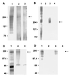
(A) Silver-stained 2 to 12% gradient SDS-PAGE gel of solubilized oocysts before (lane 1, 1.5 × 10⁶) and after (lane 2, 10 μg) electrophoretic isolation of CPC205 (arrow). Lane 3 was loaded with sample buffer to identify silver-stain artifacts. (B) Western blot recognition of isolated CPC205 (lane 3, 10 μg) (arrow) and a 205-kDa comigrating antigen in solubilized oocysts (1.5 × 10⁶, lane 1) by MAb 4D3. Lane 2 (1.5 × 10⁶ solubilized oocysts) and lane 4 (10 μg of CPC205) were probed with isotype control MAb. Molecular mass standards (Bio-Rad) are indicated on the left (myosin, 204 kDa; β-galactosidase, 121 kDa; BSA, 78 kDa). (C) Silver-stained SDS–7.5% PAGE gel of solubilized T. foetus before (lane 1, 300 μg) and after (lane 2, 10 μg) electrophoretic isolation of Tf190 (arrow). Lane 3 was loaded with sample buffer, identifying a 70-kDa silver-stain artifact. (D) Western blot recognition of isolated Tf190 (lane 3, 10 μg) (arrow) and comigrating antigen in solubilized organisms (300 μg, lane 1) by MAb 32.3B3.5. Lane 2 (300 μg of solubilized T. foetus) and lane 4 (10 μg of Tf190) were probed with isotype control MAb. Molecular mass standards (Amersham) are indicated on the left (myosin, 200 kDa; β-galactosidase, 97.4 kDa; BSA, 69 kDa; carbonic anhydrase, 46 kDa).
FIG. 3
FIG. 3
2-D Western blots of isolated CSL (6 μg) demonstrating antigen species migrating in the 1,200- to 1,400-kDa region (arrows) which are recognized by MAb 3E2 (A) or MAb 4D10 (B). Note the recognition of species 1 and 2 by 3E2 but not by 4D10. Molecular mass standards (titin, 2,450 kDa; nebulin, 770 kDa) and pI values are indicated on the left and bottom, respectively, of each panel.
FIG. 4
FIG. 4
Time-lapse video photomicroscopic depiction of sporozoite interactions with Caco-2 cells after incubation with MAb 3E2. Note that sporozoites (arrows) undergoing the CSP-like reaction (arrowheads) after treatment with MAb 3E2 fail to attach and invade. Bars, 7 μm. The time p.i. (in minutes) is indicated in each frame.
FIG. 5
FIG. 5
Time-lapse video photomicroscopic depiction of sporozoite interactions with Caco-2 cells after incubation with isotype control MAb. Note a sporozoite (arrow) probing the cell surface (0:03.30), attaching (0:05.30), invading (0:07.00), and becoming intracellular (0:08.30). Bars, 7 μm. The time p.i. (in minutes) is indicated in each frame.
FIG. 6
FIG. 6
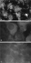
Immunofluorescence photomicrographs of Caco-2 cell monolayers incubated with IEF-isolated CSL (A), CPC205 (B), or Tf190 (C) and probed with MAbs 3E2, 4D3, or 32.3B3.5, respectively. Note the specific binding of CSL (A), indicated by immunofluorescence reactivity (arrows), and the absence of binding of control glycoproteins CPC205 (B) and Tf190 (C). Bars, 7 μm.
FIG. 7
FIG. 7
Dose-dependent reduction in Caco-2 cell permissiveness to sporozoite infection by specifically bound CSL. Caco-2 cells were incubated with MEM, or increasing amounts (0.25, 0.50, 1.0, or 2.0 μg) of IEF-isolated CSL (○), CPC205 (■), or Tf190 (▵) prior to inoculation with sporozoites. The mean numbers of intracellular stages in cells incubated with CSL were significantly lower than in those incubated with CPC205 (P < 0.002) or Tf190 (P < 0.03) at 0.50 μg, CPC205 (P < 0.004) or Tf190 (P < 0.002) at 1.0 μg, and CPC205 (P < 0.00002) or Tf190 (P < 0.00005) at 2.0 μg. The mean numbers of intracellular stages in cultures incubated with MEM (6,866 ± 126), CPC205, or Tf190 were not significantly different. Bars represent the standard deviation.
FIG. 8
FIG. 8
Kinetics of CSL binding to Caco-2 cells. (A) Dose-dependent binding of CSL to Caco-2 cells, corrected for nonspecific binding. (B) Self-displaceable binding of CSL to Caco-2 cells. The counts per minute of 125I-CSL bound in the presence of 0.13 to 4 μg of unlabeled CSL compared to that of 0.06 μg of unlabelled CSL are significantly (P < 0.02) lower. Bars represent the standard deviation (A and B). (C) Maximal binding capacity of biologically active 125I-CSL as determined from the ordinate of the y intercept for the best fit line (dashed line). (D) Saturability of CSL binding to Caco-2 cells. Bars represent the standard deviation. (E) Scatchard plot depicting the best-fit concave upward curvature line (dashed line).

Similar articles

Cited by

References

    1. Arrowood M J, Sterling C R. Isolation of Cryptosporidium oocysts and sporozoites using discontinuous sucrose and isopycnic Percoll gradients. J Parasitol. 1987;73:314–319. - PubMed
    1. Barnes D A, Bonnin A, Huang J X, Gousset L, Wu J, Gut J, Doyle P, Dubremetz J F, Ward H, Petersen C. A novel multi-domain mucin-like glycoprotein of Cryptosporidium parvum mediates invasion. Mol Biochem Parasitol. 1998;96:93–110. - PubMed
    1. Blagburn B L, Soave R. Prophylaxis and chemotherapy. In: Fayer R, editor. Cryptosporidium and cryptosporidiosis. Boca Raton, Fla: CRC Press; 1997. pp. 111–123.
    1. Brown W C, McElwain T F, Hotzel I, Suarez C E, Palmer G H. Helper T-cell epitopes encoded by the Babesia bigemina rap-1 gene family in the constant and variant domains are conserved among parasite strains. Infect Immun. 1998;66:1561–1569. - PMC - PubMed
    1. Burgess D E, McDonald C M. Analysis of adhesion and cytotoxicity of Tritrichomonas foetus to mammalian cells by use of monoclonal antibodies. Infect Immun. 1992;60:4253–4259. - PMC - PubMed

Publication types

MeSH terms