. 1999 Oct;67(10):5427-33.
doi: 10.1128/IAI.67.10.5427-5433.1999.

The intercellular adhesion (ica) locus is present in Staphylococcus aureus and is required for biofilm formation

Affiliations

The intercellular adhesion (ica) locus is present in Staphylococcus aureus and is required for biofilm formation

S E Cramton et al. Infect Immun. 1999 Oct.

Abstract

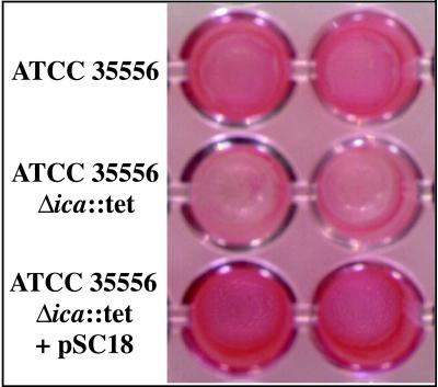
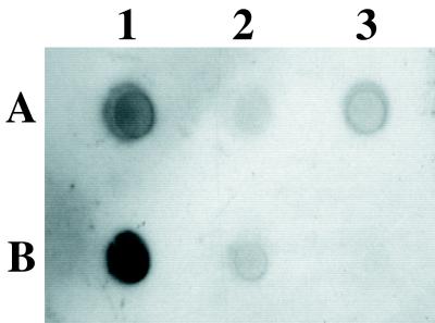
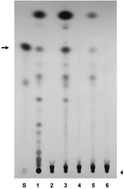
Nosocomial infections that result in the formation of biofilms on the surfaces of biomedical implants are a leading cause of sepsis and are often associated with colonization of the implants by Staphylococcus epidermidis. Biofilm formation is thought to require two sequential steps: adhesion of cells to a solid substrate followed by cell-cell adhesion, creating multiple layers of cells. Intercellular adhesion requires the polysaccharide intercellular adhesin (PIA), which is composed of linear beta-1,6-linked glucosaminylglycans and can be synthesized in vitro from UDP-N-acetylglucosamine by products of the intercellular adhesion (ica) locus. We have investigated a variety of Staphylococcus aureus strains and find that all strains tested contain the ica locus and that several can form biofilms in vitro. Sequence comparison with the S. epidermidis ica genes revealed 59 to 78% amino acid identity. Deletion of the ica locus results in a loss of the ability to form biofilms, produce PIA, or mediate N-acetylglucosaminyltransferase activity in vitro. Cross-species hybridization experiments revealed the presence of icaA in several other Staphylococcus species, suggesting that cell-cell adhesion and the potential to form biofilms is conserved within this genus.


Figures

FIG. 1
FIG. 1
Biofilm formation in S. aureus strains. The strains listed in Table 1 were grown overnight in polystyrol microtiter wells in TSB supplemented with 0.25% glucose. The cells that adhered to the plate after washing were then visualized by staining with safranin.
FIG. 2
FIG. 2
Map of the ica locus and surrounding sequence in S. aureus ATCC 35556. (A) Genomic organization of the ica locus and surrounding chromosomal region. The region between primers SA12 and SA11 was amplified by PCR and cloned into vector pBT5, creating plasmid pSC18. The solid line indicates the region sequenced and submitted to the EMBL/GenBank/DDBJ nucleotide sequence data libraries; the dashed line indicates previously published sequence (database accession no. M90693). (B) Schematic of pSC23 diagramming the knockout construct. Plasmid pSC18 was amplified by inverse PCR and primers SA14 and SA15, which deleted the sequence between the middle of icaR and the end of icaC. The tetracycline resistance cassette from pT181 (tet) was then ligated into the deleted ica gene locus.
FIG. 3
FIG. 3
Loss of biofilm formation in S. aureus ATCC 35556Δica::tet. The ica locus in S. aureus ATCC 35556 was deleted and replaced with a tetracycline resistance cassette by homologous recombination. The knockout strain, ATCC 35556Δica::tet, is unable to form a biofilm in vitro; however, the ability to form a biofilm is restored when the knockout strain is complemented with pSC18, carrying the wild type ica genes. The assay for each strain is shown in duplicate.
FIG. 4
FIG. 4
Loss of PIA production in S. aureus ATCC 35556Δica::tet. Cell surface extracts from overnight cultures of S. aureus ATCC 35556 (A1), ATCC 35556Δica::tet (A2), ATCC 35556Δica::tet carrying pSC18 (A3), S. epidermidis ATCC 35984 (RP62A) (B1), S. epidermidis O-47 (B2), and S. carnosus TM300 (B3) were treated with proteinase K, and PIA production was detected with an anti-S. epidermidis PIA antibody, showing that PIA is no longer produced in the ica knockout strain and is restored in the complemented mutant.
FIG. 5
FIG. 5
N-Acetylglucosaminyltransferase activity is mediated by the ica locus. Crude membrane extracts were incubated with radiolabeled UDP-N-acetylglucosamine, and synthesized oligomers were separated on an NH2-HPTLC plate. Lane S contains the standard, N-acetylglucosamine, alone. The remaining lanes contain products synthesized by crude membrane extracts from S. carnosus carrying pTXicaADBC, an inducible expression plasmid containing S. epidermidis icaA, icaD, icaB, and icaC (lane 1), S. carnosus harboring the vector, pTX16, alone (lane 2), S. epidermidis ATCC 35984 (RP62A), which produces PIA (lane 3), S. epidermidis 5179, a strain that does not produce PIA and does not form biofilms (lane 4), S. aureus wild-type strain ATCC 35556 (lane 5), and S. aureus ica knockout strain ATCC 35556Δica::tet (lane 6). An arrow indicates the monomer. Oligomers of increasing size are seen as a ladder-like series of spots that descend on the plate toward the origin (arrowhead) at the bottom. The smear just above the origin is unreacted UDP-N-acetylglucosamine.

Similar articles

Cited by

References

    1. Augustin J, Rosenstein R, Wieland B, Schneider U, Schnell N, Engelke G, Entian K-D, Götz F. Genetic analysis of epidermin biosynthetic genes and epidermin-negative mutants of Staphylococcus epidermidis. Eur J Biochem. 1992;204:1149–1154. - PubMed
    1. Baker A S, Schein O D. Ocular infections. In: Bisno A L, Waldvogel F A, editors. Infections associated with indwelling medical devices. 2nd ed. Washington, D.C: ASM Press; 1994. pp. 111–134.
    1. Bradford M M. A rapid and sensitive method for the quantitation of microgram quantities of protein utilizing the principle of protein-dye binding. Anal Biochem. 1976;72:248–254. - PubMed
    1. Brückner R. Gene replacement in Staphylococcus carnosus and Staphylococcus xylosus. FEMS Microbiol Lett. 1997;151:1–8. - PubMed
    1. Christensen G D, Baldassarri L, Simpson W A. Colonization of medical devices by coagulase-negative staphylococci. In: Bisno A L, Waldvogel F A, editors. Infections associated with indwelling medical devices. 2nd ed. Washington, D.C: American Society for Microbiology; 1994. pp. 45–78.

Publication types

Substances

Associated data

LinkOut - more resources