. 1999 Oct;67(10):5530-7.
doi: 10.1128/IAI.67.10.5530-5537.1999.

Pseudomonas aeruginosa induces type-III-secretion-mediated apoptosis of macrophages and epithelial cells

Affiliations

Pseudomonas aeruginosa induces type-III-secretion-mediated apoptosis of macrophages and epithelial cells

A R Hauser et al. Infect Immun. 1999 Oct.

Abstract

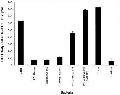
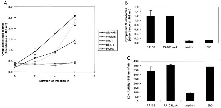
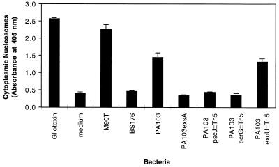
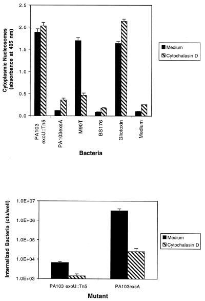
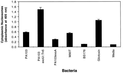
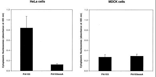
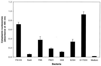
Pseudomonas aeruginosa is a gram-negative opportunistic pathogen that is cytotoxic towards a variety of eukaryotic cells. To investigate the effect of this bacterium on macrophages, we infected J774A.1 cells and primary bone-marrow-derived murine macrophages with the P. aeruginosa strain PA103 in vitro. PA103 caused type-III-secretion-dependent killing of macrophages within 2 h of infection. Only a portion of the killing required the putative cytotoxin ExoU. By three criteria, terminal deoxynucleotidyltransferase-mediated dUTP-biotin nick end labeling assays, cytoplasmic nucleosome assays, and Hoechst staining, the ExoU-independent but type-III-secretion-dependent killing exhibited features of apoptosis. Extracellular bacteria were capable of inducing apoptosis, and some laboratory and clinical isolates of P. aeruginosa induced significantly higher levels of this form of cell death than others. Interestingly, HeLa cells but not Madin-Darby canine kidney cells were susceptible to type-III-secretion-mediated apoptosis under the conditions of these assays. These findings are consistent with a model in which the P. aeruginosa type III secretion system transports at least two factors that kill macrophages: ExoU, which causes necrosis, and a second, as yet unidentified, effector protein, which induces apoptosis. Such killing may contribute to the ability of this organism to persist and disseminate within infected patients.


Figures

FIG. 1
FIG. 1
LDH release by J774A.1 cells infected with PA103 or mutants defective in type III secretion. Cells were infected for 3 h at an MOI of approximately 80. Mutants with defects in the type III secretion transcriptional activator (PA103exsA::Ω), the secretion apparatus (PA103pscJ::Tn5), or the translocation apparatus (PA103pcrG::Tn5) were noncytotoxic, whereas an exoU mutant (PA103exoU::Tn5) exhibited intermediate cytotoxic capacity. A plasmid (pAH807) containing an intact ExoU-encoding gene and its chaperone complemented the exoU mutant to a phenotype of full cytotoxicity. Error bars represent standard errors of the means for experiments performed in triplicate.
FIG. 2
FIG. 2
Apoptosis of J774A.1 cells infected with PA103. (A) PA103 caused apoptosis of J774A.1 cells in a time-dependent manner as measured by the quantitative assay ELISAPlus. Cells were infected at an MOI of approximately 160. Positive controls included gliotoxin (5 μM) and the virulent S. flexneri strain M90T. Negative controls included medium and the avirulent S. flexneri strain BS176. (B) Apoptosis-inducing capacity of PA103, PA103exsA::Ω, PA103tox::Ω, and SLO, as measured by the quantitative ELISA. Cells were infected for 6 h at an MOI of approximately 160 or exposed to SLO at a concentration of 37.5 μg/ml. (C) Cytotoxicity-inducing capacity of PA103, PA103exsA::Ω, PA103tox::Ω, and SLO, as measured by LDH release assays. Infection conditions were similar to those described in panel B. The ELISA for apoptosis is clearly capable of distinguishing necrotic cell death (SLO) from apoptotic cell death (PA103). Furthermore, exotoxin A does not contribute to PA103-induced apoptosis under the conditions of this assay. Error bars represent standard errors of the means for experiments performed in triplicate.
FIG. 3
FIG. 3
Apoptosis of J774A.1 cells infected with type III secretion mutants of PA103. J774A.1 cells were infected for 6 h at an MOI of approximately 160 and then tested for apoptosis by using the quantitative ELISA for apoptosis. Bacterial mutants with type III secretion pathway defects were associated with significantly less apoptosis than wild-type PA103. Interestingly, PA103exoU::Tn5, which has an intact secretion pathway but does not secrete the effector protein ExoU, was associated with wild-type levels of apoptosis, indicating that this factor is not necessary for induction of programmed cell death. Error bars represent standard errors of the means for experiments performed in triplicate.
FIG. 4
FIG. 4
Effect of cytochalasin D on P. aeruginosa-induced apoptosis of J774A.1 cells. (A) Quantitative ELISAs for apoptosis on J774A.1 cells infected with either PA103exoU::Tn5 (an exoU mutant that induces apoptosis) or PA103exsA::Ω (a mutant with a generalized defect in type III secretion that does not induce apoptosis). Gliotoxin (5 μM), medium, and the S. flexneri strains M90T and BS176 were used as controls. Cells were infected for 6 h at an MOI of approximately 160 in the presence or absence of 5 μg of cytochalasin D/ml. The ability of M90T to induce apoptosis was significantly inhibited by the addition of cytochalasin D, since this bacterium must be internalized to cause apoptosis. In contrast, the ability of P. aeruginosa PA103exoU::Tn5 to cause apoptosis was unaffected by cytochalasin D. (B) Effect of cytochalasin D on internalization of P. aeruginosa by J774A.1 cells. The number of internalized bacteria was significantly reduced in the presence of cytochalasin D. These results suggest that, unlike S. flexneri, extracellular P. aeruginosa causes type-III-mediated apoptosis. Error bars represent standard errors of the means for experiments performed in triplicate.
FIG. 5
FIG. 5
Quantitative ELISAs for apoptosis of primary bone-marrow-derived macrophages infected with PA103 and isogenic type III secretion mutants. Macrophages were infected for 13 h at an approximate MOI of 320 with P. aeruginosa PA103 or isogenic mutants with type III secretion defects or with S. flexneri M90T or BS176. As was the case with J774A.1 cells, apoptosis of P. aeruginosa-infected primary macrophages required an intact type III secretion pathway but not ExoU. Samples infected with PA103 exhibited significantly less apoptosis than those infected with PA103exoU::Tn5, an isogenic mutant that did not secrete ExoU, because secretion of this protein was associated with significant necrosis over the 13-h course of the experiment. Thus, fewer cells were present at the end of the assay to show signs of apoptosis. Error bars represent standard errors of the means for experiments performed in triplicate.
FIG. 6
FIG. 6
Quantitative ELISAs for apoptosis of HeLa cells (A) and MDCK cells (B) infected with wild-type PA103 or PA103exsA::Ω, a mutant with a generalized defect in type III secretion. HeLa cells were infected for 6 h at an MOI of approximately 160. MDCK cells were infected for 6 h at an MOI of approximately 40. HeLa cells were susceptible to type III-mediated apoptosis, whereas MDCK cells were not. Error bars represent standard errors of the means for experiments performed in triplicate.
FIG. 7
FIG. 7
Quantitative ELISAs for apoptosis of J774A.1 cells infected with one of several different P. aeruginosa strains. PAK, PAO1, and 388 are commonly used laboratory strains. 6294 is a hyperinvasive corneal isolate, and 617000 is a respiratory isolate. These strains were used to infect J774A.1 cells for 6 h at an MOI of approximately 160, after which cells were tested for evidence of apoptosis by using quantitative ELISAs. Strains varied markedly in their abilities to induce programmed cell death. Error bars represent standard errors of the means for experiments performed in triplicate.

Similar articles

Cited by

References

    1. Apodaca G, Bomsel M, Lindstedt R, Engel J, Frank D, Mostov K, Wiener-Kronish J. Characterization of Pseudomonas aeruginosa-induced MDCK cell injury: glycosylation defective host cells are resistant to bacterial killing. Infect Immun. 1995;63:1541–1551. - PMC - PubMed
    1. Bjorn M J, Pavlovskis O R, Thompson M R, Iglewski B H. Production of exoenzyme S during Pseudomonas aeruginosa infection in burned mice. Infect Immun. 1979;24:837–842. - PMC - PubMed
    1. Buja L M, Eigenbrodt M L, Eigenbrodt E H. Apoptosis and necrosis: basic types and mechanisms of cell death. Arch Pathol Lab Med. 1993;117:1208–1214. - PubMed
    1. Chen L M, Kone K, Galan J E. Salmonella spp. are cytotoxic for cultured macrophages. Mol Microbiol. 1996;21:1101–1115. - PubMed
    1. Coburn J, Frank D. Macrophages and epithelial cells respond differently to the Pseudomonas aeruginosa type III secretion system. Infect Immun. 1999;67:3151–3154. - PMC - PubMed

Publication types

LinkOut - more resources