Comparative Study
. 1999 Oct;181(19):5940-7.
doi: 10.1128/JB.181.19.5940-5947.1999.

Functions of two types of NADH oxidases in energy metabolism and oxidative stress of Streptococcus mutans

Affiliations
Comparative Study

Functions of two types of NADH oxidases in energy metabolism and oxidative stress of Streptococcus mutans

M Higuchi et al. J Bacteriol. 1999 Oct.

Abstract

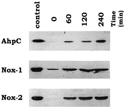
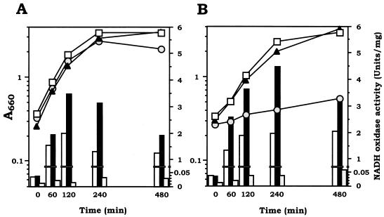
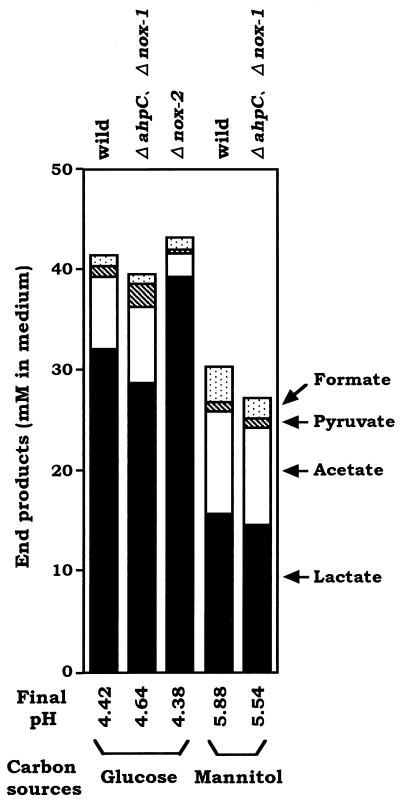
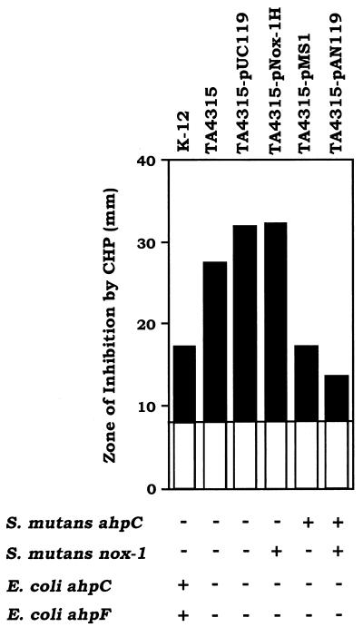
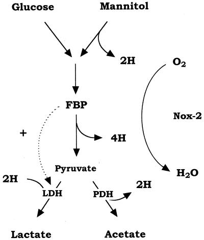
We have previously identified two distinct NADH oxidases corresponding to H(2)O(2)-forming oxidase (Nox-1) and H(2)O-forming oxidase (Nox-2) induced in Streptococcus mutans. Sequence analyses indicated a strong similarity between Nox-1 and AhpF, the flavoprotein component of Salmonella typhimurium alkyl hydroperoxide reductase; an open reading frame upstream of nox-1 also showed homology to AhpC, the direct peroxide-reducing component of S. typhimurium alkyl hydroperoxide reductase. To determine their physiological functions in S. mutans, we constructed knockout mutants of Nox-1, Nox-2, and/or the AhpC homologue; we verified that Nox-2 plays an important role in energy metabolism through the regeneration of NAD(+) but Nox-1 contributes negligibly. The Nox-2 mutant exhibited greatly reduced aerobic growth on mannitol, whereas there was no significant effect of aerobiosis on the growth on mannitol of the other strains or growth on glucose of any of the strains. Although the Nox-2 mutants grew well on glucose aerobically, the end products of glucose fermentation by the Nox-2 mutant were substantially shifted to higher ratios of lactic acid to acetic acid compared with wild-type cells. The resistance to cumene hydroperoxide of Escherichia coli TA4315 (ahpCF-defective mutant) transformed with pAN119 containing both nox-1 and ahpC genes was not only restored but enhanced relative to that of E. coli K-12 (parent strain), indicating a clear function for Nox-1 as part of an alkyl hydroperoxide reductase system in vivo in combination with AhpC. Surprisingly, the Nox-1 and/or AhpC deficiency had no effect on the sensitivity of S. mutans to cumene hydroperoxide and H(2)O(2), implying that the existence of some other antioxidant system(s) independent of Nox-1 in S. mutans compensates for the deficiency.


Figures

FIG. 1
FIG. 1
Expression of the AhpC, Nox-1, and Nox-2 proteins in S. mutans wild-type strain GS-5 before and 60, 120, and 240 min after exposure to air on TYG medium. Each protein was analyzed by immunoblotting as described in Materials and Methods. Each lane was loaded with 5 μg of protein from the corresponding extract. The leftmost lane shows the purified enzymes applied as controls (from top to bottom, 50 ng of AhpC, 100 ng of Nox-1, and 100 ng of Nox-2).
FIG. 2
FIG. 2
Aerobic growth of S. mutans wild-type strain GS-5 and the ΔahpC Δnox-1, Δnox-2, and ΔahpC Δnox-1 Δnox-2 mutants on glucose and on mannitol agar plates. (A) Colonial morphologies of wild-type (a), ΔahpC Δnox-1 (b), Δnox-2 (c), and ΔahpC Δnox-1 Δnox-2 (d) cells aerobically grown on TYG plates, incubated at 37°C for 60 h; (B) colonial morphologies of wild-type (e), ΔahpC Δnox-1 (f), Δnox-2 (g), and ΔahpC Δnox-1 Δnox-2 (h) cells aerobically grown on TYM plates, incubated at 37°C for 60 h.
FIG. 3
FIG. 3
Aerobic induction of NADH oxidase activity in S. mutans wild-type strain GS-5 and the ΔahpC Δnox-1, Δnox-2, and ΔahpC Δnox-1 Δnox-2 mutants on TYG (A) and TYM (B) media. Anaerobically grown cultures in early log phase were exposed to air and induced at 37°C by shaking under air. Growth was monitored by measuring the optical density at 660 nm of cultures of wild-type (open squares), ΔahpC Δnox-1 (closed triangles), and Δnox-2 (gray circles) cells. At the time course before and after exposure to air, the cells were harvested and NADH oxidase activity in cell extracts was assayed for wild-type (open bars), ΔahpC Δnox-1 (black bars), and Δnox-2 (shaded bars) cells. Results shown are representative of three repeated experiments.
FIG. 4
FIG. 4
Fermentation end products of S. mutans wild-type strain GS-5 and the ΔahpC, Δnox-1, and Δnox-2 mutants after 4 h of aeration in TYG (A) and TYM (B) media except that Δnox-2 was omitted. After 4 h of exposure to air, the cells were harvested and the cell-free culture media were assayed (see Materials and Methods).
FIG. 5
FIG. 5
Complementation of an ahpCF-deficient E. coli strain by S. mutans alkyl hydroperoxide reductase proteins. The zone of inhibition by 3% H2O2 or 3% cumene hydroperoxide (CHP) for E. coli TA4315 (ΔahpC ΔahpF) transformed with vector plasmid pUC119, pNox-1H containing nox-1, pMS1 containing ahpC, and pAN119 containing nox-1 and ahpC were compared with those of E. coli TA4315 as the sensitive control and E. coli K-12 (parent strain).
FIG. 6
FIG. 6
Adaptive responses against H2O2 (A) and cumene hydroperoxide (B) killing in S. mutans wild-type strain GS-5 and the ΔahpC mutant. All procedures were performed under anaerobic conditions. (A) Uninduced wild type (open circles) and ΔahpC (open triangles); H2O2-induced (60 min of treatment with 10 μM H2O2) wild type (closed circular) and ΔahpC (closed triangle). (B) Uninduced wild type (open circles) and ΔahpC (open triangles); cumene hydroperoxide-induced (60 min of treatment with 30 μM cumene hydroperoxide wild type (closed circles) and ΔahpC (closed triangles). Experiments were repeated three times, and the data shown are the means of triplicates.
FIG. 7
FIG. 7
Aerobic pathway of glucose and mannitol metabolism in S. mutans. Solid arrows indicate the flow of electron in the catabolic pathway. FBP, fructose 1,6-bisphosphate; LDH, lactate dehydrogenase; PDH, pyruvate dehydrogenase.

References

    1. Abbe K, Takahashi S, Yamada T. Involvement of oxygen-sensitive pyruvate formate-lyase in mixed-acid fermentation by Streptococcus mutans under strictly anaerobic conditions. J Bacteriol. 1982;152:175–182. - PMC - PubMed
    1. Aoki H, Shiroza T, Hayakawa M, Sato S, Kuramitsu H K. Cloning of a Streptococcus mutans glucosyltransferase gene coding for insoluble glucan synthesis. Infect Immun. 1986;53:587–594. - PMC - PubMed
    1. Bradford M M. A rapid and sensitive methods for the quantitation of microgram quantities of protein utilizing the principle of protein-dye binding. Anal Biochem. 1976;72:248–254. - PubMed
    1. Brown A T, Wittenberger C T. Fructose 1,6-diphosphate-dependent lactate dehydrogenase from a cariogenic streptococcus: purification and regulatory properties. J Bacteriol. 1972;122:1126–1135. - PMC - PubMed
    1. Carlsson J, Kujala U, Edlund M B K. Pyruvate dehydrogenase activity in Streptococcus mutans. Infect Immun. 1985;49:674–678. - PMC - PubMed

Publication types

MeSH terms

LinkOut - more resources