

doi: 10.1128/JB.181.19.6210-6213.1999.
Functional expression of hMYH, a human homolog of the Escherichia coli MutY protein
Affiliations
- PMID: 10498741
- PMCID: PMC103656
- DOI: 10.1128/JB.181.19.6210-6213.1999
Item in Clipboard
Functional expression of hMYH, a human homolog of the Escherichia coli MutY protein
J Bacteriol.
1999 Oct.
Abstract
We have previously described the hMYH cDNA and genomic clones (M. M. Slupska et al., J. Bacteriol. 178:3885-3892, 1996). Here, we report that the enzyme expressed from an hMYH cDNA clone in Escherichia coli complements the mutator phenotype in a mutY mutant and can remove A from an A. 8-hydroxydeoxyguanine mismatch and to a lesser extent can remove A from an A. G mismatch in vitro.
Figures
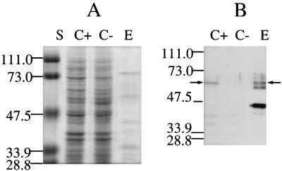
(A) Sodium dodecyl sulfate-polyacrylamide gel analysis of hMYH purified by Ni2+-agarose affinity chromatography, followed by ssDNA-cellulose chromatography (E). The proteins were separated on a 10% polyacrylamide gel in the presence of sodium dodecyl sulfate and stained with Coomassie blue. Lane S, molecular mass standards. Molecular masses, in kilodaltons, are marked on the side. Lane C+, lysate of cells of the CC104 mutY mutant containing pQE30/hMYH; lane C−, lysate of the same strain with pQE30. (B) Western blot analysis of hMYH. Proteins were separated on a 10% polyacrylamide gel, transferred onto a Hybond enhanced chemiluminescence nitrocellulose membrane (Amersham Pharmacia Inc., Piscataway, N.J.), and reacted with antibodies against the histidine tag [anti-RGS(H)4; Qiagen, Inc.]. Western blotting was performed by enhanced chemiluminescence analysis (ECL; Amersham). Arrows point to the position of full-length hMYH. Lanes C+, C−, and E are as described for panel A.
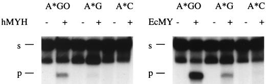
Reaction of hMYH and E. coli MutY (EcMY) with different mispair-containing 96-mer templates. Reactions were carried out for 3 h at 37°C; 3 μl of the protein preparation described in the legend to Fig. 1 and 6 ng of E. coli MutY protein were used for all templates. E. coli MutY was purified as described previously (6). The asterisks each indicate an A from a radiolabelled strand. s, substrate; p, product.
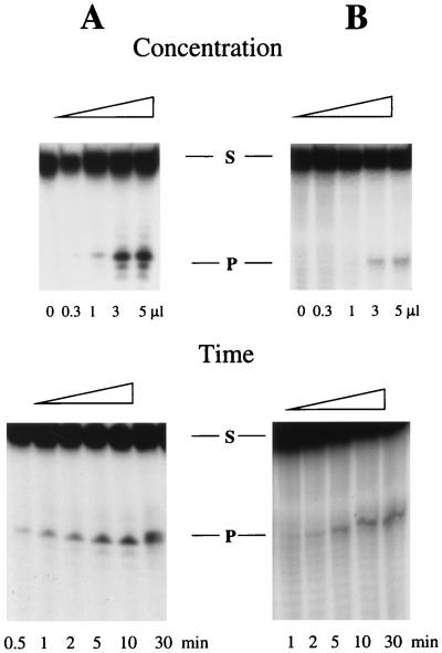
Concentration dependence and time course for the enzymatic reaction of hMYH with A · 8-oxodG placed in a 23-mer (A) and A · G placed in a 45-mer (B). Reaction conditions were as described previously (13). The sequence of the A-containing 23-mer was 5′AGAGGAAAGGAGAGAAGGGAGAG3′. The sequence of the 45-mer for the A-containing strand was 5′TTAGAGCTTGACGGGGAAAGCCAAATTCGGCGAACGTGGCGAGAA3′ (bold A marks the position of the mispaired A for both templates). In the variable-concentration experiment, reaction with A · 8-oxodG was carried out for 10 min, whereas reaction with A · G was carried out for 60 min. In the time course experiment for the A · 8-oxodG substrate, 0.3 μl of the enzyme solution was used, and for the A · G substrate, 1 μl of the enzyme was used. The A-containing oligonucleotide was radiolabelled. S and P, substrate and product, respectively.
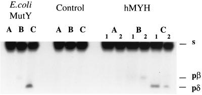
Products generated by hMYH under different conditions of heating. Reactions were stopped by freezing in a dry ice bath (lanes A), loading buffer was added and reaction mixtures were heated for 5 min at 95°C (lanes B), and panels an equal volume of 20% piperidine was added and the reaction mixture was heated for 30 min at 95°C, dried under vacuum, and dissolved in formamide loading buffer (lanes C). Products were resolved on a 15% polyacrylamide–8 M urea gel under conditions described previously (25). Numbers 1 and 2 indicate the enzyme from two different preparations of hMYH. s, migration of substrate; pβ, product of β-elimination; pδ, product of δ-elimination. Samples without enzyme were used for the control. Reactions were carried out with an A · 8-oxodG mispair placed in a 23-mer, with the A-containing strand radiolabelled, for 10 min at 37°C. The sequence of the 23-mer is in the legend to Fig. 3.
References
-
- Guan Y, Manuel R, Arvai A, Parikh S, Mol C, Miller J, Lloyd S, Tainer J. MutY catalytic core, mutant and bound adenine structures define specificity for DNA repair enzyme superfamily. Nat Struct Biol. 1998;5:1058–1064. - PubMed
-
- Lu A-L, Fawcett W P. Characterization of the recombinant MutY homolog, an adenine DNA glycosylase, from yeast Schizosaccharomyces pombe. J Biol Chem. 1998;273:25098–25105. - PubMed
Publication types
MeSH terms
Substances
Grants and funding
LinkOut - more resources
Full Text Sources
Research Materials
