. 1999 Sep;128(1):124-34.
doi: 10.1038/sj.bjp.0702775.

Endothelium-derived hyperpolarizing factor but not NO reduces smooth muscle Ca2+ during acetylcholine-induced dilation of microvessels

Affiliations

Endothelium-derived hyperpolarizing factor but not NO reduces smooth muscle Ca2+ during acetylcholine-induced dilation of microvessels

S S Bolz et al. Br J Pharmacol. 1999 Sep.

Abstract

1. We hypothesized that nitric oxide (NO) and the endothelium-dependent hyperpolarizing factor (EDHF) may dilate microvessels by different cellular mechanisms, namely Ca2+-desensitization versus decrease in intracellular free calcium. 2. Effects of acetylcholine (ACh) and the NO donors sodium nitroprusside (SNP, 0.1 - 10 micromol l(-1)) and S-Nitroso-N-acetyl-D, L-penicillamine (SNAP, 0.01 - 10 micromol l-1) on intracellular calcium ([Ca2+]i, fura 2) and vascular diameter (videomicroscopy) were studied in isolated resistance arteries from hamster gracilis muscle (194+/-12 microm) pretreated with indomethacin and norepinephrine. Membrane potential changes were determined using 1, 3-dibutylbarbituric acid trimethineoxonol (DiBAC4(3)). 3. ACh (0.1 and 1 micromol l-1)-induced dilations were associated with a [Ca2+]i decrease (by 13+/-3 and 32+/-4%) and hyperpolarization of vascular smooth muscle (VSM, by 12+/-1% at 1 micromol l-1 ACh). Nomega-nitro-L-arginine (L-NA, 30 micromol l(-1)) partially inhibited the dilation but did not affect VSM [Ca2+]i decreases or hyperpolarization. In contrast, the KCa channel inhibitors tetrabutylammonium (TBA, 1 mmol l(-1)) and charybdotoxin (ChTX, 1 micromol l(-1)) abolished the ACh-induced [Ca2+]i decrease and the hyperpolarization in VSM while a significant dilation remained (25 and 40%). This remaining dilation was abolished by L-NA. ChTX did not affect [Ca2+]i increase and hyperpolarization in endothelial cells. SNP- or SNAP-induced dilations were not associated with decreases in VSM [Ca2+]i or hyperpolarization although minor transient decreases in VSM [Ca2+]i were observed at high concentrations. 4. These data suggest that ACh-induced dilations in microvessels are predominantly mediated by a factor different from NO and PGI2, presumably EDHF. EDHF exerts dilation by activation of KCa channels and a subsequent decrease in VSM [Ca2+]i, NO dilates the microvessels in a calcium-independent manner.


Figures

Figure 1
Figure 1
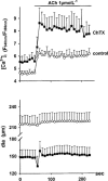
(A) Norepinephrine and acetylcholine-induced changes of diameter (dia) and intracellular calcium ([Ca2+]i) in small skeletal arteries. Small skeletal arteries were constricted with 0.3 μmol l−1 norepinephrine (NE) and subsequently stimulated with 1 μmol l−1 acetylcholine (ACh). Original recordings of changes in smooth muscle calcium ([Ca2+]i, ratio F340 nm/F380 nm) and diameter (μm) are shown in the top graphs. The box plots in the bottom panels summarize ratios and diameters under basal (‘basal'), NE-stimulated (‘NE') and ACh-stimulated (‘ACh') conditions. (Box plots represent mean±s.e.mean, n=8, *=significantly different (P<0.05) from basal value). (B) Original recording of acetylcholine-induced changes of membrane potential. Membrane potential changes were measured using the potential-sensitive dye DiBAC(4)3. Vessels were preconstricted with 1 μmol l−1 NE before ACh (1 μmol l−1) was added under control conditions (control) and in the presence of charybdotoxin (ChTX, 1 μmol l−1). Under control conditions, the fluorescence signal decreased by 12±1% after application of 1 μmol l−1 ACh, indicating hyperpolarization. This was completely inhibited in presence of ChTX.
Figure 1
Figure 1
(A) Norepinephrine and acetylcholine-induced changes of diameter (dia) and intracellular calcium ([Ca2+]i) in small skeletal arteries. Small skeletal arteries were constricted with 0.3 μmol l−1 norepinephrine (NE) and subsequently stimulated with 1 μmol l−1 acetylcholine (ACh). Original recordings of changes in smooth muscle calcium ([Ca2+]i, ratio F340 nm/F380 nm) and diameter (μm) are shown in the top graphs. The box plots in the bottom panels summarize ratios and diameters under basal (‘basal'), NE-stimulated (‘NE') and ACh-stimulated (‘ACh') conditions. (Box plots represent mean±s.e.mean, n=8, *=significantly different (P<0.05) from basal value). (B) Original recording of acetylcholine-induced changes of membrane potential. Membrane potential changes were measured using the potential-sensitive dye DiBAC(4)3. Vessels were preconstricted with 1 μmol l−1 NE before ACh (1 μmol l−1) was added under control conditions (control) and in the presence of charybdotoxin (ChTX, 1 μmol l−1). Under control conditions, the fluorescence signal decreased by 12±1% after application of 1 μmol l−1 ACh, indicating hyperpolarization. This was completely inhibited in presence of ChTX.
Figure 2
Figure 2
Effect of L-NA on ACh-induced changes of arteriolar diameter and intracellular calcium ([Ca2+]i) precontracted with 0.3 μM norepinephrine (NE). The arterial segments (n=8) were exposed to increasing concentrations of acetylcholine (ACh, from A to C) before and after treatment of the same vessels with L-NA (30 μM) and after removal of the endothelium (−E). The left-hand panel depicts diameter changes (Δ dia) while the right-hand panel shows corresponding changes of intracellular free calcium (Δ [Ca2+]i) in vascular smooth muscle. (Values represent mean±s.e.mean, arrows indicate start of ACh-application; *=significantly different (P<0.05) from control curve; n.s.=no significant difference).
Figure 3
Figure 3
Effects of TBA-pretreatment (1 mmol l−1 and additional inhibition of NO-synthase (L-NA, 30 μmol l−1 on acetylcholine (ACh)-induced changes of [Ca2+]i and diameter (n=6). 1 μmol l−1 ACh was applied to norepinephrine (NE, 0.3 μmol l−1) -preconstricted vessels. For the sake of clarity, means±s.e.mean are depicted only at intervals of 10 s. Connecting lines comprise all values (5  s intervals). Note that TBA induced rhythmic changes of [Ca2+]i in four out of six small arteries. (*=significantly different (P<0.05) from ACh under control conditions, F-test; control diameters were 114±5 μm (untreated), 109±4 μm (TBA) and 108±6 μm (TBA+L-NA). They were not significantly different).
Figure 4
Figure 4
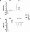
Effects of charybdotoxin (ChTX) on ACh-induced changes of [Ca2+]i, diameter (dia) and membrane potential (MP). (A) Changes in diameter and [Ca2+]i after application of ACh under control conditions, in the presence of ChTX or after combined treatment with ChTX and L-NA. Columns represent the steady state values 2 min after application of 1 μmol l−1 ACh under control conditions, after treatment with ChTX and after combined treatment with ChTX and L-NA. (n=5–6; *=significant differences to ACh effects without inhibitor(s); control diameters were 85±8 (ChTX) and 86±8 (ChTX+L-NA)). (B) Effects of ChTX on membrane potential changes in smooth muscle and endothelial cells after application of ACh. Membrane potential measurements in vascular smooth muscle and endothelial cells were performed in separate experiments. (n=4; *=significant differences to ACh effects without inhibitor(s)).
Figure 5
Figure 5
Effects of charybdotoxin (ChTX) on ACh-induced endothelial [Ca2+]i increases. ACh (1 μmol l−1) induced increases of [Ca2+]i in endothelial cells of L-NA/indomethacin-pretreated vessels (n=6). The associated dilation completely antagonized spontaneous tone. Incubation of the vessel with ChTX (1 μmol l−1, n=6) induced increases of resting endothelial [Ca2+]i (by 19%) and constricted the vessel (by 31%). Subsequent addition of ACh induced increases of [Ca2+]i as seen under control conditions but the dilation was completely inhibited.
Figure 6
Figure 6
Effect of increasing extracellular potassium ([K+]ex) concentration on [Ca2+]i and vascular diameter. The elevation of [K+]ex concentration from 4.7 to 12.5 mmol l−1 induced vasodilation (n=15) and decreased [Ca2+]i significantly (P<0.01), similar to observations with high concentrations of ACh. In contrast, further elevation of [K+]ex to 25 mmol l−1 induced vasoconstriction preceded by an increase of [Ca2+]i (P<0.01). Changes are expressed as per cent of control values (means±s.e.mean) in NE-preconstricted vessels (absolute values see text).
Figure 7
Figure 7
Dose-dependent effects of acetylcholine (ACh, (A)) and the NO donors (NOD, (B)) sodium nitroprusside (SNP) and S-Nitroso-N-acetyl-D,L-penicillamine (SNAP) on diameter and [Ca2+]i. Diameter (Δ dia) and [Ca2+]i values (Δ [Ca2+]i) represent steady state changes (per cent of control) 2 min after application of ACh or NOD to norepinephrine-preconstricted vessels. NOS-inhibition by L-NA (top panel) significantly (*=P<0.05) reduced the dilation at the lower ACh-concentration but did not affect the changes of [Ca2+]i. NOD had no effect on [Ca2+]i. (Values represent the mean±s.e.mean of seven (SNAP) or eight (ACh, SNP) experiments obtained at 2 min after application of ACh, SNAP or SNP to norepinephrine (NE, 0.3 μmol l−1)-preconstricted vessels).
Figure 8
Figure 8
Effects of the calcium channel antagonist felodipine on [Ca2+]i and vascular diameter in NE-preconstricted arteries. Top panels: Original recordings of the effect of felodipine on smooth muscle calcium ([Ca2+]i, ratio F340 nm/F380 nm) and corresponding diameter changes (μm). Bottom panels: Columns are summarizing calcium levels and diameters as obtained in nine experiments. Depicted are basal values and steady state conditions after NE as well as after application of felodipine. (*=significantly different (P<0.05) from basal value (1); for technical reasons the ratio F340 nm/F380 nm in this experimental series is higher than in the other).

References

    1. ADEAGBO A.S., HENZEL M.K. Calcium-dependent phospholipase A2 mediates the production of endothelium-derived hyperpolarizing factor in perfused rat mesentery prearteriolar bed. J. Vascular Res. 1998;35:27–35. - PubMed
    1. ANDRIANTSITOHAINA R., LAGAUD G.J.L., ANDRE A., MULLER B., STOCLET J.C. Effects of cGMP on calcium handling in ATP-stimulated rat resistance arteries. Am. J. Physiol. 1995;268:H1223–H1231. - PubMed
    1. BOLOTINA V.M., NAJIBI S., PALACINO J.J., PAGANO P.J., COHEN R.A. Nitric oxide directly activates calcium-dependent potassium channels in vascular smooth muscle. Nature. 1994;368:850–853. - PubMed
    1. BRÄUNER T., HÜLSER D.F., STRASSER R.J. Comparative measurements of membrane potentials with microelectrodes and voltage-sensitive dyes. Biochim. Biophys. Acta. 1984;771:208–216. - PubMed
    1. COHEN R.A., PLANE F., NAJIBI S., HUK I., MALINSKI T., GARLAND C.J. Nitric oxide is the mediator of both endothelium-dependent relaxation and hyperpolarization of the rabbit carotid artery. Proc. Natl. Acad. Sci. U.S.A. 1997;94:4193–4198. - PMC - PubMed

Publication types

MeSH terms

LinkOut - more resources