. 1999 Sep 20;190(6):815-26.
doi: 10.1084/jem.190.6.815.

Cleavage by granzyme B is strongly predictive of autoantigen status: implications for initiation of autoimmunity

Affiliations

Cleavage by granzyme B is strongly predictive of autoantigen status: implications for initiation of autoimmunity

L Casciola-Rosen et al. J Exp Med. .

Abstract

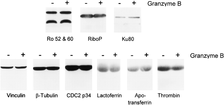
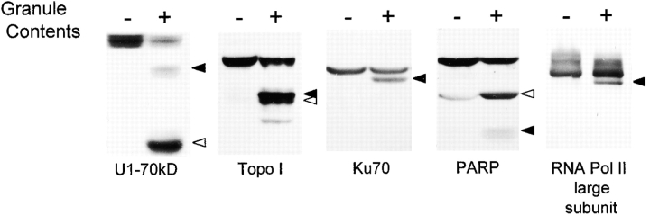
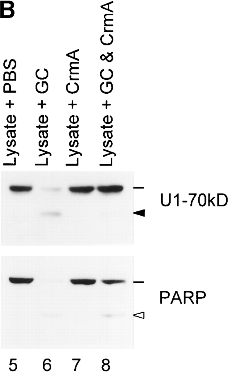
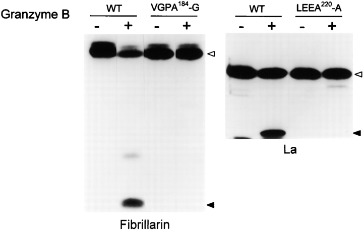
Systemic autoimmune diseases are a genetically complex, heterogeneous group of disorders in which the immune system targets a diverse but highly specific group of intracellular autoantigens. The molecules targeted are not unified by common structure, function, or distribution in control cells but become clustered and concentrated in surface blebs when cells undergo apoptosis. We show here that the majority of autoantigens targeted across the spectrum of human systemic autoimmune diseases are efficiently cleaved by granzyme B in vitro and during cytotoxic lymphocyte granule-induced death, generating unique fragments not observed during any other form of apoptosis. These molecules are not cleaved by caspase-8, although this protease has a very similar specificity to granzyme B. The granzyme B cleavage sites in autoantigens contain amino acids in the P(2) and P(3) positions that are preferred by granzyme B but are not tolerated by caspase-8. In contrast to autoantigens, nonautoantigens are either not cleaved by granzyme B or are cleaved to generate fragments identical to those formed in other forms of apoptosis. The striking ability of granzyme B to generate unique fragments is therefore an exclusive property of autoantigens and unifies the majority of molecules targeted in this spectrum of diseases. These results focus attention on the role of the cytotoxic lymphocyte granule-induced death pathway in the initiation and propagation of systemic autoimmunity.


Figures

Figure 1
Figure 1
Endogenous autoantigens are cleaved by granzyme B, generating unique fragments. (A) Gel samples were prepared from control and apoptotic (UVB-irradiated) HeLa cells and from HeLa lysate incubated in vitro with 0.8 nM purified caspase-3 and 5 mM DTT or 42 nM granzyme B and 2 mM IAA. 75-μg aliquots were electrophoresed in each gel lane, and autoantigens were detected by immunoblotting. Indicator lines denote intact antigens (Mi-2, 240 kD; PARP, 113 kD; SRP-72, 72 kD; Ki-67, 395 kD; U1-70kD, 70 kD; and topoisomerase I, 100 kD). Solid arrowheads mark granzyme B–specific fragments (Mi-2, 75, 72, and 48 kD; PARP, 110, 72, 62, and 55 kD; SRP-72, 62 kD; Ki-67, 167 and 148 kD; U1-70kD, 60 kD; topoisomerase I, 98, 75, and 72 kD). Open arrowheads denote fragments generated during apoptosis (Mi-2, 50 and 58 kD; PARP, 89 kD; SRP-72, 68 kD; Ki-67, 155, 140, 130, and 110 kD; U1-70kD, 40 kD; topoisomerase I, 70 kD). Note that the PARP autoradiogram shown was optimized for visualization of intact PARP and the 89-kD fragment; the 72- and 62-kD granzyme B–specific PARP fragments are clearly seen on longer autoradiogram exposures. (B) HeLa lysates were incubated for 1 h in the absence of protease (leftmost lane) or with the increasing amounts of granzyme B indicated. The samples were blotted with topoisomerase I antibodies. The data shows that the cleavage site that generates the 98-kD fragment from the 100-kD intact antigen (IEAD15-F; Table ) is exquisitely sensitive to granzyme B, with ∼70% of topoisomerase I cleaved to the 98-kD form after incubation with 0.42 nM granzyme B. Note that the 75- and 72-kD fragments are only generated after incubation with 10-fold higher concentrations of granzyme B.
Figure 1
Figure 1
Endogenous autoantigens are cleaved by granzyme B, generating unique fragments. (A) Gel samples were prepared from control and apoptotic (UVB-irradiated) HeLa cells and from HeLa lysate incubated in vitro with 0.8 nM purified caspase-3 and 5 mM DTT or 42 nM granzyme B and 2 mM IAA. 75-μg aliquots were electrophoresed in each gel lane, and autoantigens were detected by immunoblotting. Indicator lines denote intact antigens (Mi-2, 240 kD; PARP, 113 kD; SRP-72, 72 kD; Ki-67, 395 kD; U1-70kD, 70 kD; and topoisomerase I, 100 kD). Solid arrowheads mark granzyme B–specific fragments (Mi-2, 75, 72, and 48 kD; PARP, 110, 72, 62, and 55 kD; SRP-72, 62 kD; Ki-67, 167 and 148 kD; U1-70kD, 60 kD; topoisomerase I, 98, 75, and 72 kD). Open arrowheads denote fragments generated during apoptosis (Mi-2, 50 and 58 kD; PARP, 89 kD; SRP-72, 68 kD; Ki-67, 155, 140, 130, and 110 kD; U1-70kD, 40 kD; topoisomerase I, 70 kD). Note that the PARP autoradiogram shown was optimized for visualization of intact PARP and the 89-kD fragment; the 72- and 62-kD granzyme B–specific PARP fragments are clearly seen on longer autoradiogram exposures. (B) HeLa lysates were incubated for 1 h in the absence of protease (leftmost lane) or with the increasing amounts of granzyme B indicated. The samples were blotted with topoisomerase I antibodies. The data shows that the cleavage site that generates the 98-kD fragment from the 100-kD intact antigen (IEAD15-F; Table ) is exquisitely sensitive to granzyme B, with ∼70% of topoisomerase I cleaved to the 98-kD form after incubation with 0.42 nM granzyme B. Note that the 75- and 72-kD fragments are only generated after incubation with 10-fold higher concentrations of granzyme B.
Figure 2
Figure 2
[35S]methionine-labeled autoantigen substrates generated by in vitro transcription/translation are cleaved by purified granzyme B. [35S]methionine-labeled CENP-B, fibrillarin, PMS1, and PMS2 were incubated for 60 min at 37°C with the following amounts of caspase-3: 1 nM (PMS2) or 2.5 nM (CENP-B, fibrillarin, and PMS1). Separate incubations were performed as follows with granzyme B: 62.5 nM, 15 min (fibrillarin, PMS1); 42 nM, 60 min (CENP-B); and 28 nM, 60 min (PMS2). The samples were electrophoresed and visualized as described in Materials and Methods. Open arrowheads denote intact antigens (CENP-B, 80 kD; fibrillarin, 37 kD; PMS1, 110 kD; PMS2, 105 kD). Solid arrowheads mark the granzyme B–specific fragments (CENP-B, 58 and 40 kD; fibrillarin, 17 kD; PMS1, 50 and 60 kD; PMS2, 60, 50, and 36 kD).
Figure 3
Figure 3
Immunoprecipitated endogenous autoantigens are cleaved by purified granzyme B. [35S]methionine/cysteine-labeled endogenous histidyl tRNA synthetase, RNA polymerase II, PMScl, and alanyl tRNA synthetase were immunoprecipitated and cleaved by granzyme B as described in Materials and Methods. Solid arrowheads denote granzyme B–specific fragments (histidyl tRNA synthetase, 40 kD; RNA polymerase II large subunit, 190, 110, and 92 kD; PMScl, 85 and 74 kD; alanyl tRNA synthetase, 58 kD).
Figure 4
Figure 4
Numerous proteins are not cleaved by granzyme B. HeLa cell lysates were incubated for 60 min in the absence or presence of 42 nM granzyme B, followed by gel electrophoresis of 75-μl aliquots. The susceptibility of the autoantigens Ro (52 and 60 kD), ribosomal protein P, Ku-80, and nonautoantigens vinculin, β-tubulin, and Cdcp34 to cleavage by granzyme B was assessed by immunoblotting. Although none of these molecules was cleaved, efficient cleavage of NuMA, PARP, and U1-70kD in these lysates confirmed granzyme B activity (data not shown). The ability of granzyme B to cleave the purified nonautoantigens lactoferrin, apotransferrin, and thrombin was examined by incubating 20 μg of each substrate in the absence or presence of 30 nM granzyme B for 60 min at 37°C, followed by Coomassie blue staining of SDS-PAGE.
Figure 5
Figure 5
Granzyme B–specific autoantigen fragments are generated after in vivo incubation of intact K562 cells with YT cell GC. K562 cells were incubated in the absence (−) or presence (+) of YT cell GC for 2 h at 37°C. After terminating the reactions by adding gel buffer, 55 μg of protein was electrophoresed in each gel lane, and the samples were immunoblotted with monospecific patient sera. Solid and open arrowheads mark the positions of the granzyme B–specific and caspase-specific fragments, respectively.
Figure 6
Figure 6
Granzyme B–specific autoantigen fragments are generated in K562 cells attacked by LAK cells. LAK cells were incubated with K562 cells in the absence or presence of 100 μM Ac-DEVD-CHO 21. After terminating the reactions, the following numbers of cells were electrophoresed in each gel lane: 3 × 10⁵ LAK cells (lane 1); 105 K562 cells (lane 2); 3 × 10⁵ LAK cells plus 105 K562 cells (lanes 3 and 4). Autoantigens were detected by immunoblotting with monospecific patient sera. Solid and open arrowheads mark the positions of granzyme B–specific and caspase-specific fragments, respectively; indicator lines denote the intact antigens.
Figure 7
Figure 7
Generation of unique autoantigen fragments is abolished by Z-IETD-FMK or CrmA. Indicator lines denote the positions of intact antigens; solid and open arrowheads denote the granzyme B–specific and caspase-specific fragments, respectively. (A) LAK and K562 cells were preincubated in the absence or presence of 50 μM Z-IETD-FMK and then coincubated (E/T ratio of 3:1) for an additional 4 h 21. The samples were electrophoresed and autoantigens were blotted as described in the Fig. 6 legend. (B) GC were preincubated in the absence or presence of an equimolar amount of CrmA (amounts based on the granzyme B content of GC) for 5 min at 37°C. HeLa lysates were then incubated with these GC for a further 60 min at 37°C (0.5 μl GC per 75 μg lysate) before electrophoresis and immunoblotting.
Figure 7
Figure 7
Generation of unique autoantigen fragments is abolished by Z-IETD-FMK or CrmA. Indicator lines denote the positions of intact antigens; solid and open arrowheads denote the granzyme B–specific and caspase-specific fragments, respectively. (A) LAK and K562 cells were preincubated in the absence or presence of 50 μM Z-IETD-FMK and then coincubated (E/T ratio of 3:1) for an additional 4 h 21. The samples were electrophoresed and autoantigens were blotted as described in the Fig. 6 legend. (B) GC were preincubated in the absence or presence of an equimolar amount of CrmA (amounts based on the granzyme B content of GC) for 5 min at 37°C. HeLa lysates were then incubated with these GC for a further 60 min at 37°C (0.5 μl GC per 75 μg lysate) before electrophoresis and immunoblotting.
Figure 8
Figure 8
Granzyme B cleavage of fibrillarin and La is abolished by P1 D→A mutation. Site-directed mutation of aspartic acid residues in the P1 positions of fibrillarin (D184→A) and La (D220→A) was performed. Wild-type (WT) and mutated products were generated by in vitro transcription/translation before cleavage with granzyme B as described in Materials and Methods. Open and solid arrowheads mark the positions of intact molecules and granzyme B–specific fragments, respectively. Similar results were obtained when P1 aspartic acid residues in the granzyme B cleavage sites of U1-70kD, Mi-2, PMS1, PMS2, and topoisomerase I were mutated (data not shown).
Figure 9
Figure 9
Caspase-8 does not cleave endogenous autoantigens that are granzyme B substrates. HeLa lysate (in which endogenous caspases had been inactivated by 1 mM IAA) was incubated with 5 mM DTT in the absence (lanes 1, 4, 7, 9, 11, and 13) or presence (lanes 2, 5, 8, 10, 12, and 14) of 50 nM caspase-8 for 1 h at 37°C. In the lysates shown in lanes 3 and 6, omission of IAA permitted caspase-8 to activate endogenous effector caspases. After terminating the reactions, 75-μg amounts of sample were electrophoresed in each gel lane. Autoantigens were detected by blotting with monospecific patient sera. In lanes 1–6, open and solid arrowheads denote the SDS-PAGE migration positions of the intact antigens and the caspase-8–induced fragments, respectively. Radiolabeled PMS1 and PMS2 (generated by in vitro transcription/translation) were also not cleaved by purified caspase-8 (data not shown).

References

    1. Hardin J.A., Rahn D.R., Shen C., Lerner M.R., Wolin S.L., Rosa M.D., Steitz J.A. Antibodies from patients with connective tissue diseases bind specific subsets of cellular RNA–protein particles. J. Clin. Invest. 1982;70:141–147. - PMC - PubMed
    1. Tan E.M. Antinuclear antibodiesdiagnostic markers for autoimmune diseases and probes for cell biology. Adv. Immunol. 1989;44:93–151. - PubMed
    1. Casciola-Rosen L.A., Anhalt G., Rosen A. Autoantigens targeted in systemic lupus erythematosus are clustered in two populations of surface structures on apoptotic keratinocytes. J. Exp. Med. 1994;179:1317–1330. - PMC - PubMed
    1. Mamula M.J. The inability to process a self-peptide allows autoreactive T cells to escape tolerance. J. Exp. Med. 1993;177:567–571. - PMC - PubMed
    1. Sercarz E.E., Lehmann P.V., Ametani A., Benichou G., Miller A., Moudgil K. Dominance and crypticity of T cell antigenic determinants. Annu. Rev. Immunol. 1993;11:729–766. - PubMed

Publication types