. 1999 Sep 20;190(6):875-80.
doi: 10.1084/jem.190.6.875.

Idiopathic pulmonary alveolar proteinosis as an autoimmune disease with neutralizing antibody against granulocyte/macrophage colony-stimulating factor

Affiliations

Idiopathic pulmonary alveolar proteinosis as an autoimmune disease with neutralizing antibody against granulocyte/macrophage colony-stimulating factor

T Kitamura et al. J Exp Med. .

Abstract

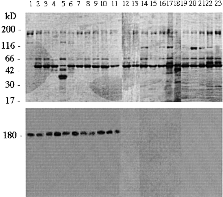
Idiopathic pulmonary alveolar proteinosis (I-PAP) is a rare disease of unknown etiology in which the alveoli fill with lipoproteinaceous material. We report here that I-PAP is an autoimmune disease with neutralizing antibody of immunoglobulin G isotype against granulocyte/macrophage colony-stimulating factor (GM-CSF). The antibody was found to be present in all specimens of bronchoalveolar lavage fluid obtained from 11 I-PAP patients but not in samples from 2 secondary PAP patients, 53 normal subjects, and 14 patients with other lung diseases. It specifically bound GM-CSF and neutralized bioactivity of the cytokine in vitro. The antibody was also found in sera from all I-PAP patients examined but not in sera from a secondary PAP patient or normal subjects, indicating that it exists systemically in I-PAP patients. As lack of GM-CSF signaling causes PAP in congenital cases and PAP-like disease in murine models, our findings strongly suggest that neutralization of GM-CSF bioactivity by the antibody causes dysfunction of alveolar macrophages, which results in reduced surfactant clearance.


Figures

Figure 1
Figure 1
Occurrence of GM-CSF binding factor in BALF from I-PAP patients. Proteins in BALF from I-PAP patients (lanes 1–11), S-PAP patients (lanes 12–13), normal subjects (lanes 14–18), and patients with other lung diseases (namely sarcoidosis, lane 19; collagen vascular lung disease, lane 20; interstitial pneumonitis, lane 21; hypersensitive pneumonitis, lane 22; and eosinophilic pneumonia, lane 23) were subjected to SDS-PAGE under nonreducing conditions, stained with Coomassie blue (top panel), and assayed with 125I–GM-CSF (bottom panel). Molecular mass markers are shown at left (kD). Radioactive 180-kD bands are seen in all I-PAP samples but not in samples from S-PAP patients, normal subjects, or patients with other lung diseases. No such band was detected in BALF from an additional 48 normal subjects or 9 patients with other lung diseases.
Figure 2
Figure 2
Characterization of GM-CSF binding factor in BALF from I-PAP patients. Molecular mass markers are shown at left in A–E (kD). (A) Protein profiles of SDS-PAGE at each purification step (stained by silver stain). Delipidated BALF (lane 1) was purified using HiTrap SP (lane 2), HiTrap Q (lane 3), Superose 12 (lane 4), RESOURCE Q (lane 5), and RESOURCE S columns (lane 6). (B) SDS-PAGE of the purified protein under nonreducing (lane 1) and reducing conditions (lane 2). Stained by silver stain. (C) Coomassie blue staining (lanes 1 and 2) and GM-CSF binding activity (lanes 3 and 4) of crude (lanes 1 and 3) and purified factor (lanes 2 and 4). (D) GM-CSF binding activity of Ig isolated using recombinant protein A column. Protein profile stained with Coomassie blue (top panel) and results of 125I–GM-CSF binding (bottom panel) are shown. Lane 0, delipidated BALF; lanes 1–9, pass-through fractions; lanes 10–14, proteins eluted from column by changing pH gradient. (E) Competition of 125I–GM-CSF binding with nonradioactive GM-CSF. Lane 1, without nonradioactive GM-CSF; lanes 2 and 3, with 50- and 500-fold concentrations of nonradioactive GM-CSF, respectively.
Figure 2
Figure 2
Characterization of GM-CSF binding factor in BALF from I-PAP patients. Molecular mass markers are shown at left in A–E (kD). (A) Protein profiles of SDS-PAGE at each purification step (stained by silver stain). Delipidated BALF (lane 1) was purified using HiTrap SP (lane 2), HiTrap Q (lane 3), Superose 12 (lane 4), RESOURCE Q (lane 5), and RESOURCE S columns (lane 6). (B) SDS-PAGE of the purified protein under nonreducing (lane 1) and reducing conditions (lane 2). Stained by silver stain. (C) Coomassie blue staining (lanes 1 and 2) and GM-CSF binding activity (lanes 3 and 4) of crude (lanes 1 and 3) and purified factor (lanes 2 and 4). (D) GM-CSF binding activity of Ig isolated using recombinant protein A column. Protein profile stained with Coomassie blue (top panel) and results of 125I–GM-CSF binding (bottom panel) are shown. Lane 0, delipidated BALF; lanes 1–9, pass-through fractions; lanes 10–14, proteins eluted from column by changing pH gradient. (E) Competition of 125I–GM-CSF binding with nonradioactive GM-CSF. Lane 1, without nonradioactive GM-CSF; lanes 2 and 3, with 50- and 500-fold concentrations of nonradioactive GM-CSF, respectively.
Figure 3
Figure 3
Inhibition of TF-1 cell growth by isolated Ig. Concentrations used: 1 ng/ml GM-CSF, 100 μg/ml delipidated BALF, 1 μg/ml isolated Ig, and 1 ng/ml IL-3.
Figure 4
Figure 4
Occurrence of antibody against GM-CSF in sera from I-PAP patients. Proteins in sera from I-PAP patients (lanes 1–5), an S-PAP patient (lane 6), and normal subjects (lanes 7–11) were subjected to SDS-PAGE under nonreducing conditions, stained with Coomassie blue (top panel), and assayed for 125I–GM-CSF binding (bottom panel). Molecular mass markers are shown at left (kD). Radioactive 180-kD bands are seen in all I-PAP samples but not in samples from the S-PAP patient and normal subjects. No such band was detected in sera from 25 additional normal subjects.

References

    1. van Golde L.M.G., Batenburg J.J., Robertson B. The pulmonary surfactant systembiochemical aspect and functional significance. Physiol. Rev. 1988;68:374–455. - PubMed
    1. Kuroki Y., Voelker D.R. Pulmonary surfactant protein. J. Biol. Chem. 1994;269:25943–25946. - PubMed
    1. Prakash U.B., Barham S.S., Carpenter H.A., Dines D.E., Marsh H.M. Pulmonary alveolar phospholipoproteinosisexperience with 34 cases and a review. Mayo Clin. Proc. 1987;62:499–518. - PubMed
    1. Ramirez R.J. Alveolar proteinosisimportance of pulmonary lavage. Am. Rev. Respir. Dis. 1971;103:666–678. - PubMed
    1. Alberti A., Luisetti M., Braschi A., Rodi G., Iotti G., Sella D., Poletti V., Benori V., Baritussio A. Bronchoalveolar lavage fluids composition in alveolar proteinosis. Early changes after therapeutic lavage. Am. J. Respir. Crit. Care Med. 1996;154:817–820. - PubMed

MeSH terms

Substances