. 1999 Sep 15;13(18):2439-48.
doi: 10.1101/gad.13.18.2439.

A zinc-binding site in the largest subunit of DNA-dependent RNA polymerase is involved in enzyme assembly

Affiliations

A zinc-binding site in the largest subunit of DNA-dependent RNA polymerase is involved in enzyme assembly

D Markov et al. Genes Dev. .

Abstract

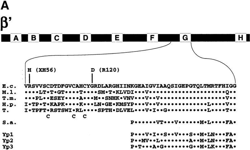
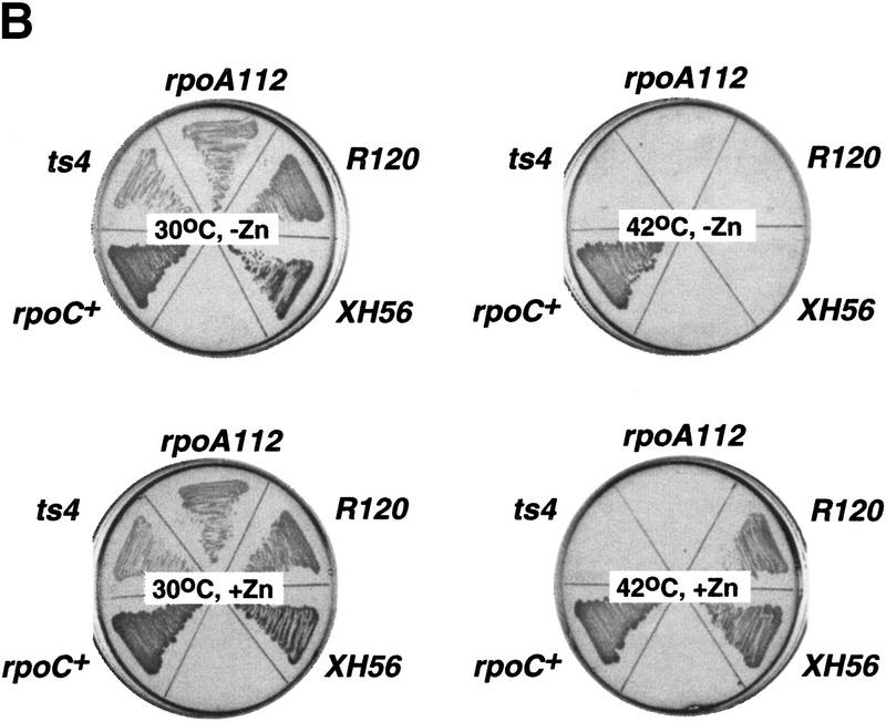
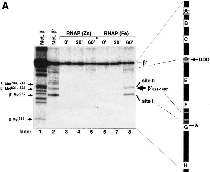
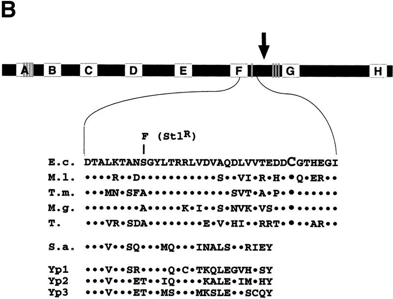
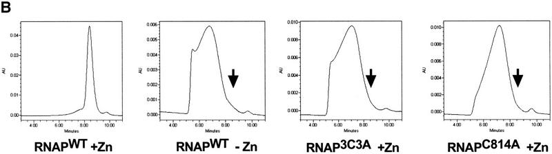
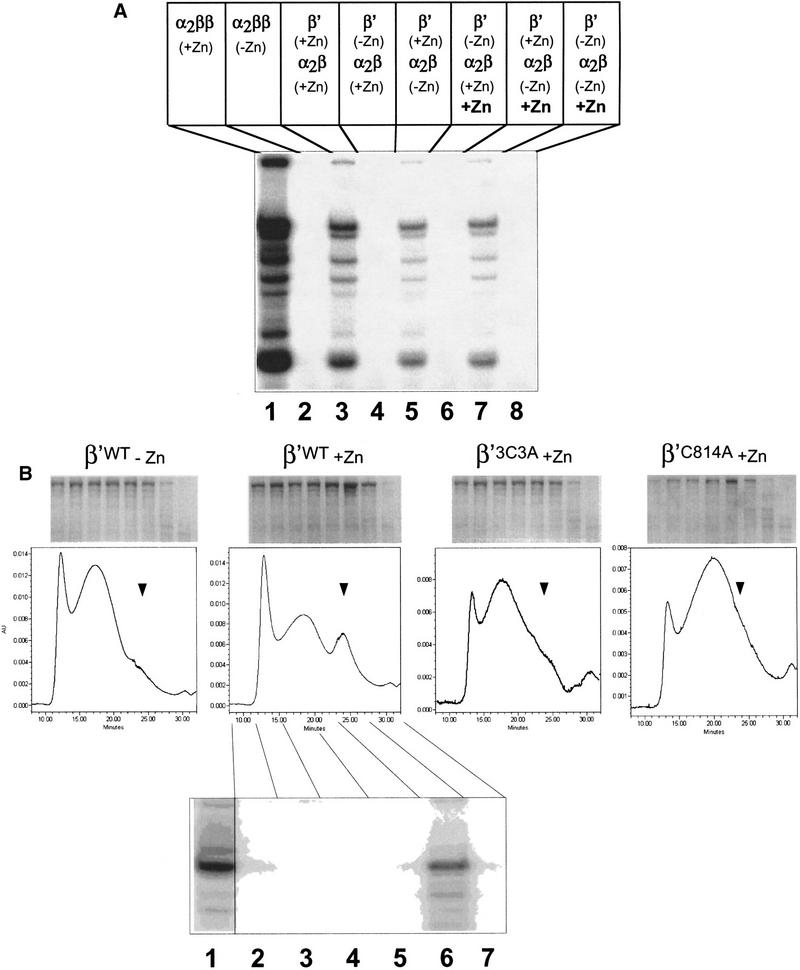
All multisubunit DNA-dependent RNA polymerases (RNAP) are zinc metalloenzymes, and at least two zinc atoms are present per enzyme molecule. RNAP residues involved in zinc binding and the functional role of zinc ions in the transcription mechanism or RNAP structure are unknown. Here, we locate four cysteine residues in the Escherichia coli RNAP largest subunit, beta', that coordinate one of the two zinc ions tightly associated with the enzyme. In the absence of zinc, or when zinc binding is prevented by mutation, the in vitro-assembled RNAP retains the proper subunit stoichiometry but is not functional. We demonstrate that zinc acts as a molecular chaperone, converting denatured beta' into a compact conformation that productively associates with other RNAP subunits. The beta' residues coordinating zinc are conserved throughout eubacteria and chloroplasts, but are absent from homologs from eukaryotes and archaea. Thus, the involvement of zinc in the RNAP assembly may be a unique feature of eubacterial-type enzymes.


Figures

Figure 1
Figure 1
The R120 and XH56 rpoC assembly mutations are suppressed by zinc. (A) Genetic context of the R120 and XH56 mutations. The bar at the top represents the 1407-amino-acid β′ subunit from E. coli. The lettered boxes indicate evolutionarily conserved segments. E. coli segment G is expanded underneath and is aligned to the corresponding segments from M. leprae (M.l.), T. maritima (T.m.), H. pylori (H.p.), and tobacco chloroplasts (T), as well as S. acidocaldarius (S.a.), and yeast RNAP I, II, and III (Yp1, Yp2, and Yp3, respectively). The XH56 and R120 mutations are indicated above the E. coli sequence; the three cysteines conserved within the eubacterial lineage are indicated below the tobacco sequence. (B) The overnight growth of the indicated E. coli strains on LB plates with or without 100 μm ZnCl2 at 30°C and 42°C.
Figure 1
Figure 1
The R120 and XH56 rpoC assembly mutations are suppressed by zinc. (A) Genetic context of the R120 and XH56 mutations. The bar at the top represents the 1407-amino-acid β′ subunit from E. coli. The lettered boxes indicate evolutionarily conserved segments. E. coli segment G is expanded underneath and is aligned to the corresponding segments from M. leprae (M.l.), T. maritima (T.m.), H. pylori (H.p.), and tobacco chloroplasts (T), as well as S. acidocaldarius (S.a.), and yeast RNAP I, II, and III (Yp1, Yp2, and Yp3, respectively). The XH56 and R120 mutations are indicated above the E. coli sequence; the three cysteines conserved within the eubacterial lineage are indicated below the tobacco sequence. (B) The overnight growth of the indicated E. coli strains on LB plates with or without 100 μm ZnCl2 at 30°C and 42°C.
Figure 2
Figure 2
Localized radical cleavage of wild-type RNAP. (A) RNAPs containing zinc or iron were affinity labeled in β′ segment G using the T7 A2 promoter-containing DNA fragment as a template and treated with 0.03% H2O2 and 1 mm sodium ascorbate for times indicated. The reaction products were resolved on a 10% SDS-polyacrylamide gel and visualized by autoradiography. (Lanes 1,2) Marker lanes generated by treating the RNAP affinity labeled at β Lys-1065 (lane 1) or β′ conserved segment G (lane 2) with CNBr under single-hit conditions. The mobility of the recombinant β′821–1407 fragment is indicated by an arrow at the right of the gel. The bar at right represents E. coli β′. The affinity-labeling site in segment G is shown. The three catalytic Asp residues in segment D, the four evolutionarily conserved Cys residues in segment A, as well as the cysteines close to cleavage sites I and II are shown as shaded lines. (B) Genetic context of the β′ iron cleavage site II (see Fig. 1A legend for details). Cys-814 is highlighted by larger font size; an E. coli mutation S793F that results in streptolydigin-resistant RNAP (Severinov et al. 1995) is shown above the E. coli sequence. (↓) The position of natural split site in archaea.
Figure 2
Figure 2
Localized radical cleavage of wild-type RNAP. (A) RNAPs containing zinc or iron were affinity labeled in β′ segment G using the T7 A2 promoter-containing DNA fragment as a template and treated with 0.03% H2O2 and 1 mm sodium ascorbate for times indicated. The reaction products were resolved on a 10% SDS-polyacrylamide gel and visualized by autoradiography. (Lanes 1,2) Marker lanes generated by treating the RNAP affinity labeled at β Lys-1065 (lane 1) or β′ conserved segment G (lane 2) with CNBr under single-hit conditions. The mobility of the recombinant β′821–1407 fragment is indicated by an arrow at the right of the gel. The bar at right represents E. coli β′. The affinity-labeling site in segment G is shown. The three catalytic Asp residues in segment D, the four evolutionarily conserved Cys residues in segment A, as well as the cysteines close to cleavage sites I and II are shown as shaded lines. (B) Genetic context of the β′ iron cleavage site II (see Fig. 1A legend for details). Cys-814 is highlighted by larger font size; an E. coli mutation S793F that results in streptolydigin-resistant RNAP (Severinov et al. 1995) is shown above the E. coli sequence. (↓) The position of natural split site in archaea.
Figure 3
Figure 3
In vitro transcription by 3C3A RNAP. (A) RNAP3C3A is temperature resistant in vitro. RNAP3C3A was affinity purified from 397C cells harboring the pCYB2β′3C3A plasmid and used to transcribe the T7 A1 promoter-containing DNA fragment at the indicated temperatures. Reaction products were separated on a denaturing 20% polyacrylamide gel and revealed by autoradiography. (B) RNAP3C3A is assembly defective in vitro. RNAP3C3A and RNAPWT were subjected to a denaturation/renaturation cycle by the addition of 6 m guanidine-HCl, followed by dialysis into transcription buffer. The samples were then assayed in a steady-state transcription assay, using the T7 A1 promoter as a template. Reaction products were analyzed as in A. (C) Localized radical cleavage of iron-containing RNAP3C3A. RNAPs containing plasmid-borne, His6-tagged wild-type or 3C3A β′ were affinity purified from 397C E. coli cells grown in the presence of iron and in the absence of zinc. Affinity labeling, hydroxy-radical cleavage, and reaction-product analyses were performed as described in the legend to Fig. 2.
Figure 3
Figure 3
In vitro transcription by 3C3A RNAP. (A) RNAP3C3A is temperature resistant in vitro. RNAP3C3A was affinity purified from 397C cells harboring the pCYB2β′3C3A plasmid and used to transcribe the T7 A1 promoter-containing DNA fragment at the indicated temperatures. Reaction products were separated on a denaturing 20% polyacrylamide gel and revealed by autoradiography. (B) RNAP3C3A is assembly defective in vitro. RNAP3C3A and RNAPWT were subjected to a denaturation/renaturation cycle by the addition of 6 m guanidine-HCl, followed by dialysis into transcription buffer. The samples were then assayed in a steady-state transcription assay, using the T7 A1 promoter as a template. Reaction products were analyzed as in A. (C) Localized radical cleavage of iron-containing RNAP3C3A. RNAPs containing plasmid-borne, His6-tagged wild-type or 3C3A β′ were affinity purified from 397C E. coli cells grown in the presence of iron and in the absence of zinc. Affinity labeling, hydroxy-radical cleavage, and reaction-product analyses were performed as described in the legend to Fig. 2.
Figure 4
Figure 4
Zn2+ is not important for interactions of β′ with other RNAP subunits. (A) Binding of α2β to immobilized His6–β‘ studied by the Ni2+-coimmobilization assay. The α subunit was renatured with β, σ, and His6–β′WT in the presence or in the absence of 10 μm ZnCl2 (lanes 15 and 610, respectively), or with His6–β′3C3A (lanes 1115) or His6–β′C814A (lanes 1620) in the presence of ZnCl2, loaded onto Ni2+–NTA–agarose beads (L), and the unbound protein (or flowthrough) removed (F). The beads were then washed with buffer containing 5 mm imidazole (W1) and 25 mm imidazole (W2), and the bound proteins were eluted with buffer containing 100 mm imidazole (E). The protein fractions were analyzed by SDS-PAGE. Control experiments indicated that when His6–β‘ was omitted from the reconstitution mixture, no nonspecific binding of β and α to the Ni2+–NTA–agarose beads occurred (data not shown; Wang et al. 1997). (B) Abnormal chromatographic behavior of RNAP assembled in the absence of zinc. Proteins eluted from Ni2+–NTA beads (lanes 5,10,15,20 in A) were loaded on a SEC-400 gel-filtration column.
Figure 4
Figure 4
Zn2+ is not important for interactions of β′ with other RNAP subunits. (A) Binding of α2β to immobilized His6–β‘ studied by the Ni2+-coimmobilization assay. The α subunit was renatured with β, σ, and His6–β′WT in the presence or in the absence of 10 μm ZnCl2 (lanes 15 and 610, respectively), or with His6–β′3C3A (lanes 1115) or His6–β′C814A (lanes 1620) in the presence of ZnCl2, loaded onto Ni2+–NTA–agarose beads (L), and the unbound protein (or flowthrough) removed (F). The beads were then washed with buffer containing 5 mm imidazole (W1) and 25 mm imidazole (W2), and the bound proteins were eluted with buffer containing 100 mm imidazole (E). The protein fractions were analyzed by SDS-PAGE. Control experiments indicated that when His6–β‘ was omitted from the reconstitution mixture, no nonspecific binding of β and α to the Ni2+–NTA–agarose beads occurred (data not shown; Wang et al. 1997). (B) Abnormal chromatographic behavior of RNAP assembled in the absence of zinc. Proteins eluted from Ni2+–NTA beads (lanes 5,10,15,20 in A) were loaded on a SEC-400 gel-filtration column.
Figure 5
Figure 5
The β′ subunit becomes assembly competent in the presence of zinc ions. (A) The β′ subunit or a mixture of the α and β subunits (labeled α2β) were renatured separately either in the presence or in the absence of 10 μm ZnCl2. Proteins were then dialyzed in a buffer without zinc, combined as indicated, and incubated for 15 min at 30°C with the recombinant σ70 subunit to reconstitute RNAP either in the presence (lanes 68) or in the absence (lanes 15) of 10 μm ZnCl2. RNAP activity was then assayed using DNA fragment containing the T7 A1 promoter (see Fig. 3A legend). (Lanes 1,2) Controls, where standard RNAP reconstitution reactions containing α, β, and β′ were carried out either in the presence (lane 1) or in the absence (lane 2) of zinc. (B) Chromatographic analysis of the β′ subunits individually renatured in the presence or in the absence of zinc. The indicated β′ subunits were individually renatured and analyzed on an SEC-400 column attached to HPLC. Fractions were collected and analyzed by 8% SDS-PAGE (top). The gel at the bottom shows the results of in vitro transcription reaction using the indicated chromatographic fractions of wild-type β′ renatured in the presence of zinc. Fractions were supplemented with α2β and σ and assayed as in A.

References

    1. Allison LA, Moyle M, Shales M, Ingles CJ. Extensive homology among the largest subunits of eukaryotic and prokaryotic RNA polymerases. Cell. 1985;42:599–610. - PubMed
    1. Bartolomei MS, Corden JL. Localization of an alpha-amanitin resistance mutation in the gene encoding the largest subunit of mouse RNA polymerase II. Mol Cell Biol. 1987;7:586–594. - PMC - PubMed
    1. Borukhov S, Goldfarb A. Recombinant Escherichia coli RNA polymerase: Purification of individually overexpressed subunits and in vitro assembly. Protein Expr Purif. 1993;4:503–511. - PubMed
    1. Borukhov S, Severinov K, Kashlev M, Lebedev A, Bass I, Rowland GC, Lim PP, Glass RE, Nikiforov V, Goldfarb A. Mapping of trypsin cleavage and antibody-binding sites and delineation of a dispensable domain in the beta subunit of Escherichia coli RNA polymerase. J Biol Chem. 1991;266:23921–23926. - PubMed
    1. Chatterji D, Wu FY-H. Selective substitution in vitro of an intrinsic zinc of Escherichia coli RNA polymerase with various divalent metals. Biochemistry. 1982;21:4651–4656. - PubMed

MeSH terms

LinkOut - more resources