. 1999 Sep 28;96(20):10992-9.
doi: 10.1073/pnas.96.20.10992.

Sonic hedgehog protein signals not as a hydrolytic enzyme but as an apparent ligand for patched

Affiliations

Sonic hedgehog protein signals not as a hydrolytic enzyme but as an apparent ligand for patched

N Fuse et al. Proc Natl Acad Sci U S A. .

Abstract

The amino-terminal signaling domain of the Sonic hedgehog secreted protein (Shh-N), which derives from the Shh precursor through an autoprocessing reaction mediated by the carboxyl-terminal domain, executes multiple functions in embryonic tissue patterning, including induction of ventral and suppression of dorsal cell types in the developing neural tube. An apparent catalytic site within Shh-N is suggested by structural homology to a bacterial carboxypeptidase. We demonstrate here that alteration of residues presumed to be critical for a hydrolytic activity does not cause a loss of inductive activity, thus ruling out catalysis by Shh-N as a requirement for signaling. We favor the alternative, that Shh-N functions primarily as a ligand for the putative receptor Patched (Ptc). This possibility is supported by new evidence for direct binding of Shh-N to Ptc and by a strong correlation between the affinity of Ptc-binding and the signaling potency of Shh-N protein variants carrying alterations of conserved residues in a particular region of the protein surface. These results together suggest that direct Shh-N binding to Ptc is a critical event in transduction of the Shh-N signal.


Figures
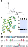
Figure 1
Figure 1
A possible catalytic site in Shh-N. (A) Model for an apparent zinc hydrolase catalytic site derived from the crystal structure of Shh-N (23). Glu-177 and His-135 residues are presumed to be essential for catalysis (see text), and His-141, Asp-148, and His-183 coordinate the Zn2+ ion. (B) Superimposed alpha-carbon traces of Shh-N (yellow) and d,d-carboxypeptidase from Streptomyces albus (green). The portion of these proteins displaying structural homology is drawn, with the Zn2+ ions shown as blue spheres. Residues within the structurally homologous portion of Shh-N that are altered in SC (four of six) and SD (two of three) (see text) are located in structurally diverged loops and are highlighted in red. (C) Coomassie blue staining of purified recombinant wild type (WT) and E177A (EA), H135A (HA) and double mutant (EH) Shh-N proteins resolved in SDS/PAGE (15%); molecular mass markers are indicated at left (kDa). (D) Structure-based alignment of amino acid sequence from the portions of mouse Shh (mSHH) and Streptomyces albus d,d-carboxypeptidase (DD-C) shown in B. The residues involved in zinc coordination or hydrogen bonding of the water molecule are shown in dark blue, and other conserved residues are in light blue. Target sites for mutagenesis are indicated in green (for zinc hydrolase mutants) or red (for SC and SD mutants, see below).
Figure 2
Figure 2
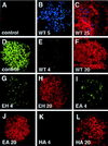
Signaling activities of Shh-N zinc hydrolase mutants. (A–C) Chicken intermediate neural plate explants double stained for expression of the motor neuron marker Islet-1(blue) and the floor plate marker HNF-3β (red). No Islet-1- or HNF-3β-positive cells were observed in control explants (A), whereas 5 nM (B) and 25 nM (C) concentrations of wild-type Shh-N induced expression of Islet-1 and HNF-3β, respectively. (D–L) Neural plate explants double stained with antibodies against a dorsal marker Pax-7 (green) and the floor plate marker HNF-3β (red). Explants cultured with medium only (D) express Pax-7 but not HNF-3β. Wild-type Shh-N protein fully repressed expression of Pax-7 at 4 nM (E) and uniformly induced HNF-3β in all cells at 20 nM (F). The EH and EA mutant proteins repressed Pax-7 at 4 nM (G, I, respectively), albeit somewhat less efficiently, and were able to uniformly induce HNF-3β expression at 20 nM (H and J, respectively). The H135A (HA) mutant protein was indistinguishable from wild type (K, at 4 nM and L, at 20 nM). Images were captured using a ×40 objective.
Figure 3
Figure 3
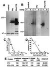
Direct binding of Shh-N to Ptc. (A) Ptc and Ptc-CTD expression in stably transfected cloned cell lines. Cell lysates were prepared from stable EcR-293 cell lines carrying pIND(Sp) (empty vector control), pIND(Sp)-Ptc, or pIND(Sp)-Ptc-CTD, and proteins were fractionated by SDS/PAGE (6%) followed by blotting and detection with anti-Ptc antibody (Santa Cruz Biotechnology). Two bands (dots) were detected in lysates from Ptc or from Ptc-CTD cells, but not from control cells. (B) Crosslinking of 32P-labeled Shh-N protein to Ptc and Ptc-CTD. EcR-293 cells expressing Ptc and Ptc-CTD were incubated with 32P-Shh-N protein in absence (−) or presence (+) of a 100-fold excess of unlabeled Shh-N protein and then crosslinked. Cell lysates were subjected to SDS/PAGE (6%) and crosslinked products detected by autoradiography. Autoradiographic images for control and Ptc and for Ptc-CTD are presented at distinct contrast settings to highlight the crosslinked species. Migration of marker proteins (in kDa) is shown at left. (C, D) Scatchard analysis of the high-affinity component of 32P-Shh-N binding to EcR-293 cells expressing Ptc (C) or Ptc-CTD (D). (E) Summary of predicted molecular masses of Ptc and Ptc-CTD, experimental values estimated from Western blotting (A), and apparent masses of crosslinked products (B). Experimental values are the average of several independent determinations. Also shown are estimates of the binding coefficients of Shh-N for Ptc and for Ptc-CTD, and estimates of the number of binding sites per cell.
Figure 4
Figure 4
Alteration of Shh-N surface residues. (A) Ribbon diagram and (B, C) surface representations of Shh-N. B is shown in the same orientation as A, but C is rotated 180° about a vertical axis relative to A and B. Surface-exposed evolutionarily conserved residues that were selected for alteration cluster into four major regions: SA (blue), SB (green), SC (red), and SD (yellow). Residues mutagenized are indicated in Table 1. (D) Coomassie blue stain of an SDS/PAGE separation of purified mutant Shh-N proteins. SE, SF, and SG denote proteins with distinct subsets of the altered residues in SC (see Table 1). Migration of molecular mass markers indicated at left (in kDa). Figs. 1B and 4A made with molscript (36); Fig. 4 B and C were made with grasp (37).
Figure 5
Figure 5
Signaling activities of Shh-N proteins with altered surface residues. Neural plate explants stained for expression of Pax-7 (green) and HNF-3β (red). Explants were cultured in the presence of the indicated proteins at the indicated concentrations (nM). SA and SB proteins are as active as wild-type Shh-N, because they repress expression of Pax-7 at 4 nM and induce expression of HNF-3β at 20 nM. The SD protein is slightly less active than wild-type protein, and the SC mutant protein is completely inactive. The SE and SG proteins display reduced activity, and the SF protein is even less active. Results are summarized in Table 1. Images were captured using a ×40 objective.
Figure 6
Figure 6
Binding of altered Shh-N proteins to Ptc. (A and B) Competition by altered proteins for binding of 32P-Shh-N to EcR-293 cells expressing Ptc-CTD. Binding of 32P-Shh-N in the presence of each altered protein at various concentrations is normalized to the total value of 32P-Shh-N bound (approximately 35% of input) in the absence of competitor. The SC mutant, inactive in signaling, also fails to compete for binding to Ptc-CTD. The SE, SF, and SG proteins with intermediate levels of signaling activity, displayed intermediate levels of competition for binding to Ptc-CTD. Data are summarized in Table 1. (C) Signaling activity as a function of Ptc affinity. On the basis of neural plate signaling assays (Figs. 2, 5; Table 1), protein concentrations required for Pax-7 repression are plotted as a function of Ptc-binding affinity. The protein concentrations are plotted as ranges centered about the concentrations presented in Table 1. Note that there is an excellent correlation between Ptc binding and activity in Pax-7 repression. The zinc hydrolase mutants EA, HA, and EH (Table 1) also corroborate this correlation but are omitted for clarity.
Figure 7
Figure 7
Reactivity of 5E1 antibody with altered Shh-N proteins. After immunoprecipitation (IP) with the 5E1 monoclonal antibody, altered proteins were detected by Western blotting using a polyclonal antibody. The starting (input) and precipitated (IP) material are shown for each protein. (A) The wild-type, SA, SB, and SD altered proteins were precipitated well by the 5E1 antibody, but the SC protein was not. (B) SE, SF, and SG displayed intermediate reactivity with the 5E1 antibody.

References

    1. Perrimon N. Cell. 1995;80:517–520. - PubMed
    1. Hammerschmidt M, Brook A, McMahon A P. Trends Genet. 1997;13:14–21. - PubMed
    1. Goodrich L V, Scott M P. Neuron. 1998;21:1243–1257. - PubMed
    1. Chiang C, Litingtung Y, Lee E, Young K, Corden J L, Westphal H, Beachy P. Nature (London) 1996;383:407–413. - PubMed
    1. Beachy P A, Cooper M K, Young K E, von Kessler D P, Park W J, Hall T M, Leahy D J, Porter J A. Cold Spring Harbor Symp Quant Biol. 1997;62:191–204. - PubMed

Publication types