. 1999 Sep 28;96(20):11008-14.
doi: 10.1073/pnas.96.20.11008.

Kinetic stability as a mechanism for protease longevity

Affiliations

Kinetic stability as a mechanism for protease longevity

E L Cunningham et al. Proc Natl Acad Sci U S A. .

Abstract

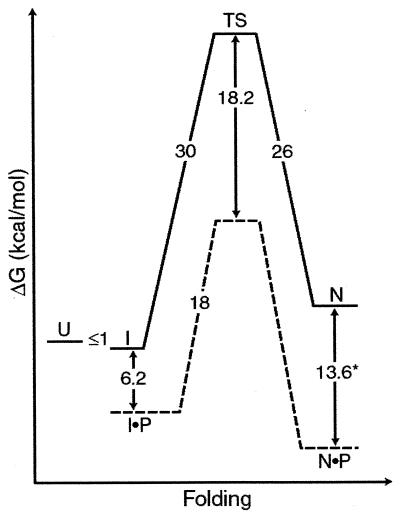
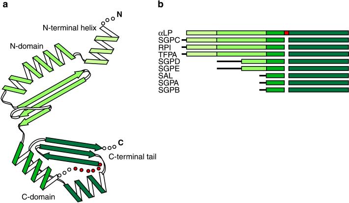
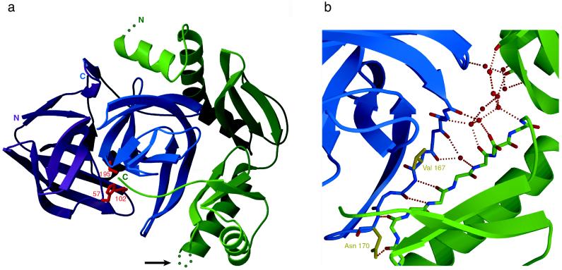
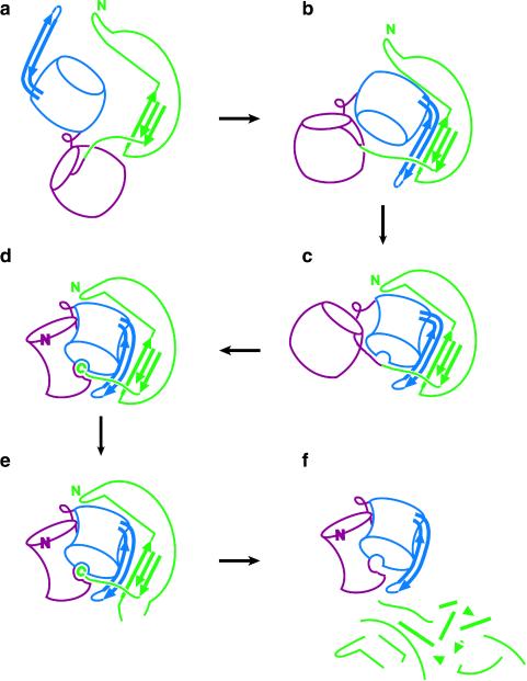
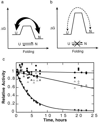
The folding of the extracellular serine protease, alpha-lytic protease (alphaLP; EC 3.4.21.12) reveals a novel mechanism for stability that appears to lead to a longer functional lifetime for the protease. For alphaLP, stability is based not on thermodynamics, but on kinetics. Whereas this has required the coevolution of a pro region to facilitate folding, the result has been the optimization of native-state properties independent of their consequences on thermodynamic stability. Structural and mutational data lead to a model for catalysis of folding in which the pro region binds to a conserved beta-hairpin in the alphaLP C-terminal domain, stabilizing the folding transition state and the native state. The pro region is then proteolytically degraded, leaving the active alphaLP trapped in a metastable conformation. This metastability appears to be a consequence of pressure to evolve properties of the native state, including a large, highly cooperative barrier to unfolding, and extreme rigidity, that reduce susceptibility to proteolytic degradation. In a test of survival under highly proteolytic conditions, homologous mammalian proteases that have not evolved kinetic stability are much more rapidly degraded than alphaLP. Kinetic stability as a means to longevity is likely to be a mechanism conserved among the majority of extracellular bacterial pro-proteases and may emerge as a general strategy for intracellular eukaryotic proteases subject to harsh conditions as well.


Figures

Figure 1
Figure 1
Free-energy diagram of αLP folding with and without its pro region at 4°C. In the absence its pro region (P), unfolded αLP (U) spontaneously folds to a molten globule-like intermediate (I), which proceeds at an extremely slow rate to N through a high-energy folding TS. The addition of pro region provides a catalyzed folding pathway (denoted by dashed lines) that lowers the high folding barrier and results in a thermodynamically stable inhibition complex N·P. ∗ indicates measurement at 25°C. (Modified from ref. .)
Figure 2
Figure 2
(a) Topology of Pro as described in the text. A disordered loop in the Pro C domain is shown in red. (b) Schematic of primary sequence alignments of pro regions from nine bacterial serine proteases. Alignments were determined by using the αLP Pro structure as a guide. Regions of sequence homology correspond to specific secondary structures in the Pro structure, with the Pro C-terminal domain being the most conserved region. N-terminal sequences lacking homology are depicted by thin black lines. αLP, Lysobacter enzymogenes αLP (17); SGPC, Streptomyces griseus protease C (18); RPI, Rarobacter faecitabitus protease I (19); SGPD, S. griseus protease D (20); SGPE, S. griseus protease E (21); TFPA, Themomonaspora fusca serine protease (22); SAL, Streptomyces lividans protease (23); SGPA, S. griseus protease A (24); SGPB, S. griseus protease B (24).
Figure 3
Figure 3
(a) Ribbon diagram of the Pro⋅N complex structure. The αLP N and C domains are colored magenta and blue respectively, with the side chains of the catalytic triad shown in red (His-57, Asp-102 and Ser-195; chymotrypsin numbering). Illustrated in green, bound Pro inserts its C-terminal tail into the protease active site. A disordered loop in the Pro C-terminal domain, indicated by an arrow, presents a likely secondary protease cleavage site, leading to the release of active αLP from the inhibitory complex. (b) Detail of the hydrated Pro⋅N interface. A gap between Pro (green) and the αLP C domain (blue) is filled by ordered water molecules which are shown as red spheres. Some of these waters mediate hydrogen bonds (dashed orange lines) between the αLP β-hairpin and the Pro three-stranded β-sheet that form the shared five-stranded β-sheet of the Pro⋅N interface. Residues in the αLP β-hairpin that affect formation of the initial Pro⋅I Michaelis complex (Ile-167 and Asn-170) are displayed in yellow. Figures are modified from figures 2b and 3b of ref. .
Figure 4
Figure 4
Proposed model of Pro-catalyzed folding of αLP. (a) The pro domain of the Pro–αLP precursor folds, while the protease N and C domains remain separated and expanded. (b) The three-stranded β-sheet of the Pro C domain pairs with the solvent-exposed β-hairpin of the αLP C domain forming a continuous five-stranded β-sheet. (c) Substrate-like binding of the Pro–αLP junction to the nascent active site positions the β-hairpin and leads to the structuring of the αLP C domain. (d) The αLP N domain folds on docking with the αLP C domain to complete the protease active site, which can then process the Pro–αLP junction. The Pro C-terminal tail remains bound to the active site in this inhibitory complex while the new αLP N terminus repositions to its native conformation. (e) Intermolecular cleavage of secondary cleavage sites by αLP or other exogenous proteases leads to the f, eventual degradation of Pro and release of active, mature αLP. Color scheme as in Fig. 3a. Figure is modified from figure 4 of ref. .
Figure 5
Figure 5
Advantages of kinetic stability. (a) A typical thermodynamically stable protein without a large barrier samples fully and partially unfolded states, making it susceptible to proteolysis. (b) A kinetically stable protein only rarely samples these unfolded states, making it much more resistant to proteolysis. In the case of αLP, the native state is less stable than the unfolded states; however, kinetic stability does not require a metastable native state. (c) αLP (●) is more resistant to proteolysis than either trypsin (▵) or chymotrypsin (♦). αLP (purified as described in ref. 25), trypsin (TPCK-treated, Worthington), and chymotrypsin (TLCK-treated, Worthington) (6.5 μM each) were mixed in 10 mM CaCl2, 50 mM Mops (pH 7.0) at 37°C. Aliquots were removed over time, and the survival of the individual proteases was measured based on their activities, which could be distinguished given their nonoverlapping specificities for different substrates (succinyl-Ala-Pro-Ala-pNA, succinyl-Ala-Ala-Pro-Arg-pNA, succinyl-Ala-Ala-Pro-Leu-pNA, used for αLP, trypsin, and chymotrypsin, respectively, all at 1 mM in 100 mM Tris, pH 8). Whereas αLP activity decreases at a rate of less than (600 hr)−1, chymotrypsin and trypsin are inactivated with rates of (4 hr)−1 and (60 hr)−1, respectively.

References

    1. Baker D, Shiau A K, Agard D A. Curr Opin Cell Biol. 1993;5:966–970. - PubMed
    1. Brayer G D, Delbaere L T J, James M N G. J Mol Biol. 1979;131:743–775. - PubMed
    1. Silen J L, Frank D, Fujishige A, Bone R, Agard D A. J Bacteriol. 1989;171:1320–1325. - PMC - PubMed
    1. Silen J L, Agard D A. Nature (London) 1989;341:462–464. - PubMed
    1. Baker D, Sohl J L, Agard D A. Nature (London) 1992;356:263–265. - PubMed

Publication types

Substances