Review
. 1999 Sep 28;96(20):11041-8.
doi: 10.1073/pnas.96.20.11041.

A proteolytic pathway that controls the cholesterol content of membranes, cells, and blood

Affiliations
Review

A proteolytic pathway that controls the cholesterol content of membranes, cells, and blood

M S Brown et al. Proc Natl Acad Sci U S A. .

Abstract

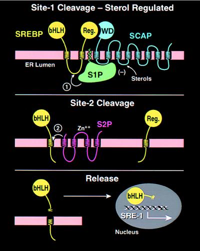
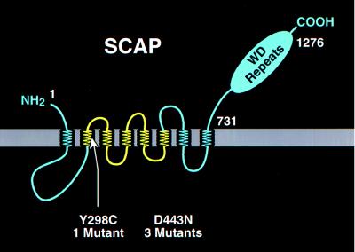
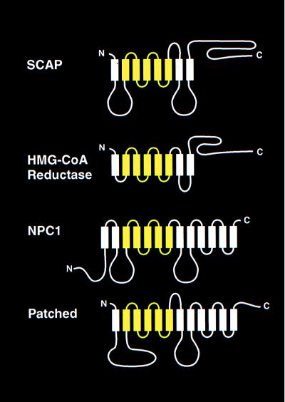
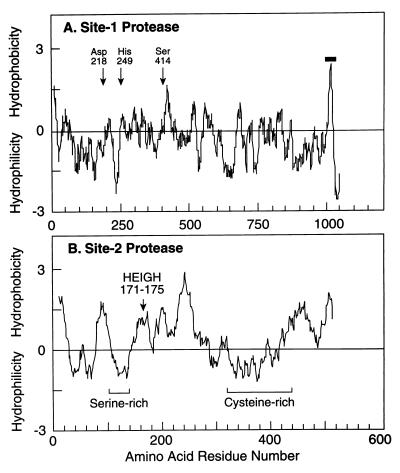
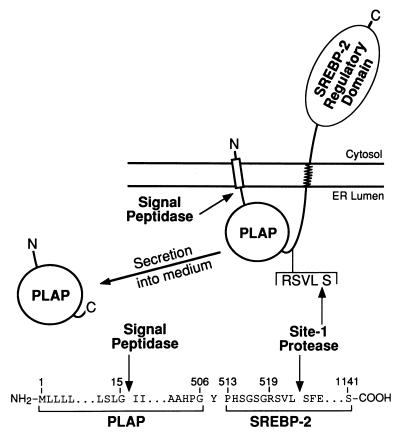
The integrity of cell membranes is maintained by a balance between the amount of cholesterol and the amounts of unsaturated and saturated fatty acids in phospholipids. This balance is maintained by membrane-bound transcription factors called sterol regulatory element-binding proteins (SREBPs) that activate genes encoding enzymes of cholesterol and fatty acid biosynthesis. To enhance transcription, the active NH(2)-terminal domains of SREBPs are released from endoplasmic reticulum membranes by two sequential cleavages. The first is catalyzed by Site-1 protease (S1P), a membrane-bound subtilisin-related serine protease that cleaves the hydrophilic loop of SREBP that projects into the endoplasmic reticulum lumen. The second cleavage, at Site-2, requires the action of S2P, a hydrophobic protein that appears to be a zinc metalloprotease. This cleavage is unusual because it occurs within a membrane-spanning domain of SREBP. Sterols block SREBP processing by inhibiting S1P. This response is mediated by SREBP cleavage-activating protein (SCAP), a regulatory protein that activates S1P and also serves as a sterol sensor, losing its activity when sterols overaccumulate in cells. These regulated proteolytic cleavage reactions are ultimately responsible for controlling the level of cholesterol in membranes, cells, and blood.


Figures

Figure 1
Figure 1
Model for the sterol-mediated proteolytic release of SREBPs from membranes. (Top) Release is initiated by Site-1 protease (S1P), a sterol-regulated protease that recognizes the SCAP/SREBP complex and cleaves SREBP in the luminal loop between two membrane-spanning sequences. SCAP allows Site-1 cleavage to be activated when cells are deprived of sterols, and it inhibits this process when sterols are abundant. (Middle) Once the two halves of SREBP are separated, a second protease, Site-2 protease (S2P), cleaves the NH2-terminal bHLH-Zip domain of SREBP at a site located within the membrane-spanning region. (Bottom) After the second cleavage, the NH2-terminal bHLH-Zip domain leaves the membrane, carrying three hydrophobic residues at its COOH-terminus. The protein enters the nucleus, where it activates target genes controlling lipid synthesis and uptake.
Figure 2
Figure 2
Membrane topology of SCAP, showing the location of two point mutations that produce a sterol-resistant phenotype in mutant cells. The yellow region denotes the putative sterol-sensing domain of SCAP.
Figure 3
Figure 3
Membrane proteins that contain sterol-sensing domains. The identified proteins are Chinese hamster SCAP (1,276 amino acids), Chinese hamster HMG-CoA reductase (887 amino acids), mouse Niemann-Pick type C1 (NPC1) (1,278 amino acids), and mouse Patched (1,434 amino acids). The sterol-sensing domains of these proteins, denoted in yellow, correspond to the following residues: SCAP, amino acids 280–446; HMG-CoA reductase, amino acids 57–224; NPC1, amino acids 617–691; and Patched, amino acids 420–589. The sequence alignments of the four sterol-sensing domains are published in Fig. 2 of ref. .
Figure 4
Figure 4
Hydropathy plots of hamster Site-1 protease (A) and human Site-2 protease (B). The residue-specific hydropathy index was calculated over a window of 20 residues by the method of Kyte and Doolittle (60) as described (44, 51). For Site-1 protease, arrows denote the three amino acids of S1P that correspond to the catalytic triad for subtilisin-like serine proteases. For Site-2 protease, the arrow denotes the sequence in S2P corresponding to the consensus HEXXH pentapeptide metal binding site for zinc metalloproteases. The one transmembrane sequence in S1P is denoted by the horizontal bar. The serine- and cysteine-rich regions in S2P are indicated.
Figure 5
Figure 5
Proteolytic processing and secretion of the PLAP/BP2 fusion protein used for the complementation cloning of S1P. The details of the construction of the plasmid encoding this fusion protein are described in ref. . In brief, the plasmid was generated by fusing the sequence encoding the signal peptide and soluble catalytic domain of human placental alkaline phosphatase (amino acids 1–506) with the sequence encoding amino acids 513–1,141 of human SREBP-2. Secretion of the catalytic domain of PLAP requires cleavage by signal peptidase and Site-1 protease. [Figure reproduced with permission from ref. (Copyright 1998, Cell Press).])

References

    1. Devaux P F. Curr Opin Struct Biol. 1993;3:489–494.
    1. Simons K, Ikonen E. Nature (London) 1997;387:569–572. - PubMed
    1. Anderson R G W. Annu Rev Biochem. 1998;67:199–225. - PubMed
    1. Brown M S, Goldstein J L. Cell. 1997;89:331–340. - PubMed
    1. Hua X, Wu J, Goldstein J L, Brown M S, Hobbs H H. Genomics. 1995;25:667–673. - PubMed

Publication types

MeSH terms