. 1999 Sep 28;96(20):11134-9.
doi: 10.1073/pnas.96.20.11134.

Purification and DNA binding properties of the ataxia-telangiectasia gene product ATM

Affiliations

Purification and DNA binding properties of the ataxia-telangiectasia gene product ATM

G C Smith et al. Proc Natl Acad Sci U S A. .

Abstract

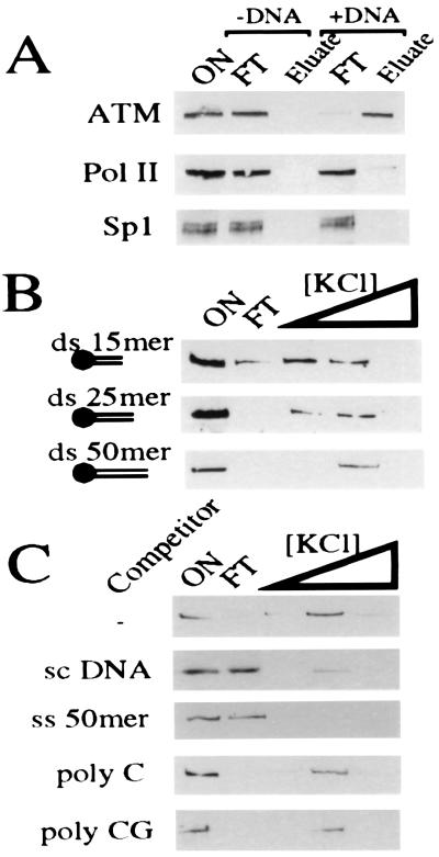
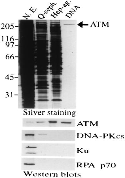
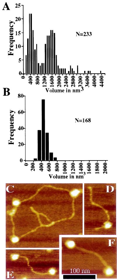
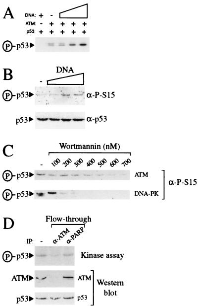
The human neurodegenerative and cancer predisposition condition ataxia-telangiectasia is characterized at the cellular level by radiosensitivity, chromosomal instability, and impaired induction of ionizing radiation-induced cell cycle checkpoint controls. Recent work has revealed that the gene defective in ataxia-telangiectasia, termed ATM, encodes an approximately 350-kDa polypeptide, ATM, that is a member of the phosphatidylinositol 3-kinase family. We show that ATM binds DNA and exploit this to purify ATM to near homogeneity. Atomic force microscopy reveals that ATM exists in two populations, with sizes consistent with monomeric and tetrameric states. Atomic force microscopy analyses also show that ATM binds preferentially to DNA ends. This property is similar to that displayed by the DNA-dependent protein kinase catalytic subunit, a phosphatidylinositol 3-kinase family member that functions in DNA damage detection in conjunction with the DNA end-binding protein Ku. Furthermore, purified ATM contains a kinase activity that phosphorylates serine-15 of p53 in a DNA-stimulated manner. These results provide a biochemical assay system for ATM, support genetic data indicating distinct roles for DNA-dependent protein kinase and ATM, and suggest how ATM may signal the presence of DNA damage to p53 and other downstream effectors.


Figures

Figure 1
Figure 1
ATM binds to DNA. (A) HeLa nuclear extract (ON) was bound to either streptavidin iron oxide beads (−DNA) or streptavidin iron oxide beads bearing a 50-mer ds DNA oligonucleotide (+DNA). After extensive washing, ATM was eluted in 500 mM KCl. Eluted proteins were subjected to 7% SDS/PAGE and ATM visualized by Western blotting using ATM.B antiserum. Equivalent fractions were probed for Sp1 and RNA polymerase II (Pol II). (B) ATM binding to DNA oligonucleotides depends on length. ATM-enriched extract (ON) was bound to streptavidin iron oxide beads attached to dsDNA of various sizes (15, 25, or 50 bp). After extensive washing, ATM was eluted sequentially with 100, 250, and 500 mM KCl. Eluates were analyzed as in A. (C) ATM is competed effectively by an excess of ssDNA and supercoiled DNA but not by poly-C or poly-CG. Washing, elution, and ATM detection was as above.
Figure 2
Figure 2
Purification of ATM to essential homogeneity. Equivalent volumes (5 μl) of HeLa cell nuclear extract (50 μg protein) or pooled fractions after sequential Q-Sepharose, Heparin-agarose, or DNA affinity chromatography were subjected to 8% SDS/PAGE and proteins visualized by silver staining (Upper). Fractions also were subjected to Western blot analysis (Lower) using antibodies raised against ATM. Filters then were stripped and reprobed by using antisera raised against DNA-PKcs, Ku70 plus Ku80 (Ku), or the 70-kDa subunit of replication protein A, as indicated.
Figure 3
Figure 3
Analysis of ATM by atomic force microscopy. (A) The volume of ATM particles adsorbed to freshly cleaved micas was calculated from atomic force micrographs. Two distinct populations were observed in the sample at 200 mM KCl. Peaks in the volume distribution are apparent at ≈400 and ≈1,600 nm3. (B) The volume of free DNA-PKcs was measured as a control for volumetric analysis of the ATM particle. DNA-PKcs displays a single peak in the volume distribution with an average particle volume of 479 nm3. (CF) Height-encoded atomic force micrographs of ATM in association with DNA. (C) ATM particles bound to supercoiled pBluescript plasmid. (D) A 660-bp linear DNA with blunt ends is bound by an ATM particle at an internal site. (E and F) The majority of linear DNAs bound by ATM are associated with the protein through an interaction involving a DNA end. (Bar = 100 nm.)
Figure 4
Figure 4
Purified ATM contains DNA-stimulatable p53 kinase activity. (A) ATM preparations phosphorylate p53 in a DNA-stimulated manner. Kinase reactions containing γ[32P]-ATP were performed either in the absence of ATM and presence of 50 ng BamHI-digested pG13CAT plasmid DNA or in the presence of ATM preparations in the absence (−) or presence of 0.5, 5, or 50 ng of BamHI-digested pG13CAT plasmid DNA. All reactions contained recombinant p53 as a substrate. Phosphorylated p53 was detected by autoradiography. (B) ATM preparations phosphorylate p53 at serine-15 in a DNA-stimulatable manner. Kinase reactions containing ATM using p53 as a substrate were performed either in the absence (−) or presence of 1, 5, or 10 ng of sheared calf thymus genomic DNA. Serine-15 phosphorylation was detected by Western blotting with phosphospecific antibodies raised against p53 phosphorylated at serine-15. (C) ATM-associated kinase activity is inhibited by wortmannin. Kinase reactions using either DNA-PK or ATM preparations and recombinant p53 as a substrate were performed in the presence of 50 ng of BamHI-digested pG13CAT plasmid DNA. Reactions were preincubated either in the absence (−) or presence of increasing concentrations of wortmannin for 30 min before the addition of ATP. Serine-15 phosphorylation was detected as in B. (D) Immunodepletion of kinase activity from ATM preparations using ATM antibody. ATM preparations were incubated in either the absence (−) or presence of ATM or poly(ADP-ribose) polymerase monoclonal antibodies as indicated. After immunodepletion, ATM preparations were used in kinase assays in the presence of γ[32P]-ATP, BamHI-digested pG13CAT plasmid DNA, and recombinant p53. Phosphorylation of p53 was monitored by autoradiography. Amounts of ATM and p53 in kinase reactions were monitored by Western blotting with ATM and p53 antibody, respectively.

References

    1. Hoekstra M F. Curr Opin Genet Dev. 1997;7:170–175. - PubMed
    1. Lavin M F, Shiloh Y. Annu Rev Immunol. 1997;15:177–202. - PubMed
    1. Rotman G, Shiloh Y. Hum Mol Genet. 1998;7:1555–1563. - PubMed
    1. Kastan M B, Zhan Q, El-Deiry W S, Carrier F, Jacks T, Walsh W V, Plunkett B S, Vogelstein B, Fornace A J J. Cell. 1992;71:587–597. - PubMed
    1. Khanna K K, Lavin M F. Oncogene. 1993;12:3307–3312. - PubMed

Publication types