. 1999 Sep 28;96(20):11241-6.
doi: 10.1073/pnas.96.20.11241.

Circular permutation and receptor insertion within green fluorescent proteins

Affiliations

Circular permutation and receptor insertion within green fluorescent proteins

G S Baird et al. Proc Natl Acad Sci U S A. .

Abstract

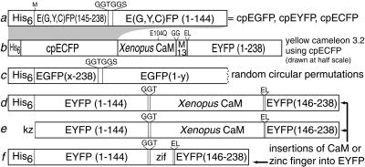
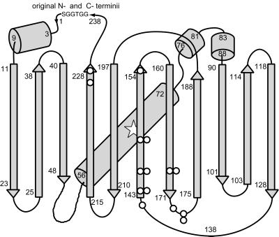
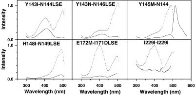
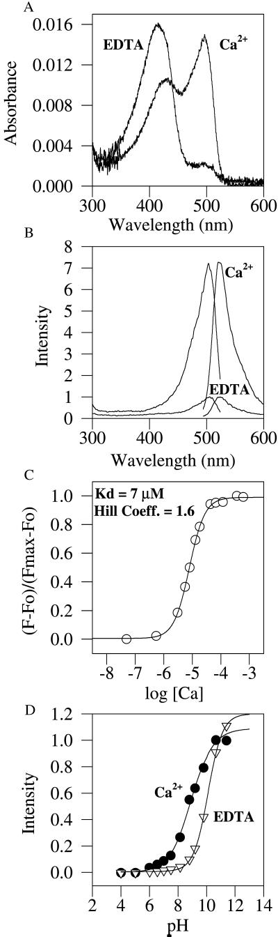
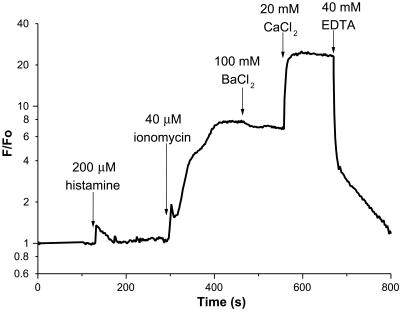
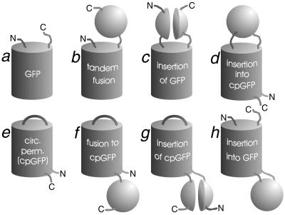
Many areas of biology and biotechnology have been revolutionized by the ability to label proteins genetically by fusion to the Aequorea green fluorescent protein (GFP). In previous fusions, the GFP has been treated as an indivisible entity, usually appended to the amino or carboxyl terminus of the host protein, occasionally inserted within the host sequence. The tightly interwoven, three-dimensional structure and intricate posttranslational self-modification required for chromophore formation would suggest that major rearrangements or insertions within GFP would prevent fluorescence. However, we now show that several rearrangements of GFPs, in which the amino and carboxyl portions are interchanged and rejoined with a short spacer connecting the original termini, still become fluorescent. These circular permutations have altered pKa values and orientations of the chromophore with respect to a fusion partner. Furthermore, certain locations within GFP tolerate insertion of entire proteins, and conformational changes in the insert can have profound effects on the fluorescence. For example, insertions of calmodulin or a zinc finger domain in place of Tyr-145 of a yellow mutant (enhanced yellow fluorescent protein) of GFP result in indicator proteins whose fluorescence can be enhanced severalfold upon metal binding. The calmodulin graft into enhanced yellow fluorescent protein can monitor cytosolic Ca(2+) in single mammalian cells. The tolerance of GFPs for circular permutations and insertions shows the folding process is surprisingly robust and offers a new strategy for creating genetically encodable, physiological indicators.


Figures

Figure 1
Figure 1
Schematic structures of major new constructs. (a) Designed circular permutations of EGFP, EYFP, and ECFP starting at Y145M. His6 indicates the polyhistidine tag MRGSHHHHHHGMASMTGGQQMGRDLYDDDDKDP. Linkers and substitutions are shown above the main sequence. (b) Yellow cameleon 3.2 (YC3.2) incorporating cpECFP instead of ECFP. This sequence is drawn at half the scale of all the other constructs. M13 is the CaM-binding peptide derived from skeletal muscle myosin light chain kinase (7). (c) Random circular permutations of EGFP. The successful values of x and y are shown in Table 1. (d and e) Insertions of CaM in place of Y145 of EYFP as expressed in bacteria (d) for in vitro purification or in HeLa cells (e) for in situ monitoring of cytosolic Ca2+. kz, Kozak sequence (10) for optimal translation initiation. (f) Insertion of a zinc finger (zif), residues 334–362 of zif268 (8), in place of Y145 of EYFP.
Figure 2
Figure 2
Schematic drawing of the overall fold of GFP (12) modified to show starting points of fluorescent circular permutations (○), the linker (GGTGGS) connecting the original N and C termini, and the approximate location of the chromophore (open star, residues 65–67). Locations with two circles indicate where circular permutations with two different ending amino acids were isolated (Table 1).
Figure 3
Figure 3
Excitation spectra of selected circular permutations of EGFP. Dotted lines (upper traces in each panel) are spectra taken at pH 8; solid lines (lower traces) are at pH 6 for the same concentration of protein. Spectra for different mutants were recorded with varying protein concentrations and gains, so amplitudes cannot be compared across permutations. The permutations are denoted by their starting and ending amino acids as in Table 1. An emission spectrum for Y145M–N144 is included in Upper Right; emission spectra for all the circular permutations were essentially identical in shape and wavelength.
Figure 4
Figure 4
In vitro characterization of EYFP–calmodulin insert protein. (A) Absorbance spectra with EDTA and zero Ca2+, or with saturating Ca2+, both at pH 7.5 and the same protein concentration. (B) Fluorescence excitation and emission spectra obtained under the same conditions. (C) Titration at pH 7.5 with Ca2+ buffers, plotting fractional conversion to the Ca2+-bound state vs. the logarithm of the free-Ca2+ concentration in molar units. (D) Titrations of normalized fluorescence vs. pH with EDTA and zero Ca2+, or with saturating Ca2+. The midpoints of the titration curves are 10.1 in EDTA and 8.9 in excess Ca2+.
Figure 5
Figure 5
Fluorescence of a typical HeLa cell expressing cytosolic EYFP–calmodulin insert as a function of time after successive additions of reagents. The fluorescence normalized by the initial fluorescence F0 is plotted on a logarithmic scale.
Figure 6
Figure 6
Topologies of GFP, cpGFP, and chimeras with other proteins. The other proteins are depicted schematically as spheres, when their sequences remain contiguous, or as paired hemispheres, when GFP or cpGFP is inserted within them. N and C denote amino and carboxyl termini of the full protein or chimera. Tandem fusions b and f arbitrarily show the carboxyl terminus of the GFP or cpGFP fused to the amino terminus of the other protein, but also are intended to encompass the opposite order. Topologies a, b, and c were already known; examples of e, f, and h are demonstrated in this paper.

References

    1. Tsien R Y. Annu Rev Biochem. 1998;67:509–544. - PubMed
    1. Siegel M S, Isacoff E Y. Neuron. 1997;19:735–741. - PubMed
    1. Biondi R M, Baehler P J, Reymond C D, Veron M. Nucleic Acids Res. 1998;26:4946–4952. - PMC - PubMed
    1. Abedi M R, Caponigro G, Kamb A. Nucleic Acids Res. 1998;26:623–630. - PMC - PubMed
    1. Heinemann U, Hahn M. Prog Biophys Mol Biol. 1995;64:121–143. - PubMed

Publication types

LinkOut - more resources