. 1999 Sep 28;96(20):11329-34.
doi: 10.1073/pnas.96.20.11329.

Galectin-7 overexpression is associated with the apoptotic process in UVB-induced sunburn keratinocytes

Affiliations

Galectin-7 overexpression is associated with the apoptotic process in UVB-induced sunburn keratinocytes

F Bernerd et al. Proc Natl Acad Sci U S A. .

Abstract

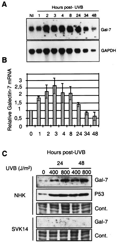
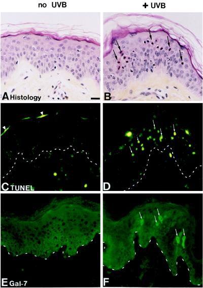
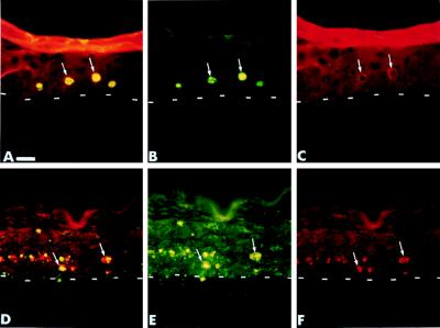
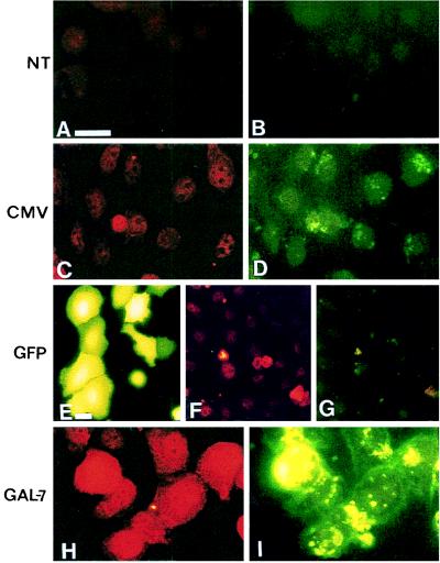
Galectin-7 is a beta-galactoside binding protein specifically expressed in stratified epithelia and notably in epidermis, but barely detectable in epidermal tumors and absent from squamous carcinoma cell lines. Galectin-7 gene is an early transcriptional target of the tumor suppressor protein P53 [Polyak, K., Xia, Y., Zweier, J., Kinzler, K. & Vogelstein, B. (1997) Nature (London) 389, 300-305]. Because p53 transcriptional activity is increased by genotoxic stresses we have examined the possible effects of ultraviolet radiations (UVB) on galectin-7 expression in epidermal keratinocytes. The amounts of galectin-7 mRNA and protein are increased rapidly after UVB irradiation of epidermal keratinocytes. The increase of galectin-7 is parallel to P53 stabilization. UVB irradiation of skin reconstructed in vitro and of human skin ex vivo demonstrates that galectin-7 overexpression is associated with sunburn/apoptotic keratinocytes. Transfection of a galectin-7 expression vector results in a significant increase in terminal deoxynucleotidyltransferase-mediated UTP end labeling-positive keratinocytes. The present findings demonstrate a keratinocyte-specific protein involved in the UV-induced apoptosis, an essential process in the maintenance of epidermal homeostasis.


Figures

Figure 1
Figure 1
UVB irradiation increases the amount of galectin-7 mRNA and proteins. (A) Normal human epidermal keratinocytes were UVB-irradiated (500 J/m2) and incubated for the indicated times (hours post-UVB) before RNA extraction and Northern blotting. A significant increase of galectin-7 mRNA relative to glyceraldehyde-3-phosphate dehydrogenase (GAPDH) mRNA is observed after UVB irradiation. (B) Scanning quantitation of blots after normalization of galectin-7 values to those of GAPDH. The average relative increase (×2.7) of the galectin-7 mRNA occurs 3–4 hr after irradiation and decreases below the initial value 48 hr after UVB irradiation. (C) Normal human epidermal keratinocytes (NHK) and SVK14 keratinocytes (SVK14) were UVB-irradiated at the doses indicated (400 or 800 J/m2) and incubated for 24 and 48 hr as indicated. Proteins then were prepared and analyzed by Western blot. Both UVB doses result in the increase of galectin-7 and P53 in NHK. These increases were more important after 800 J/m2 UVB. In SVK14 keratinocytes, galectin-7 was neither detected before or after UVB irradiation. Cont. shows a loading control (i. e. Coomassie blue staning of a portion of the gel) after transfer.
Figure 2
Figure 2
Galectin-7 expression is increased in sunburn keratinocytes formed in normal human skin ex vivo after UVB exposure. Normal human skin on grids was sham-irradiated (A, C, and E) or exposed to UVB 750 J/m2 (B, D, and F). Twenty-four hours after irradiation, samples were processed for histology (A and B), TUNEL reaction (C and D), and galectin-7 immunostaining (E and F). Histological examination reveals that UVB irradiation induces the formation of numerous sunburn cells within the suprabasal compartment of the epidermis (B, arrows). In sham-irradiated skin, rare positive nuclei restricted to the terminally differentiated keratinocytes are detected by the TUNEL reaction (C, arrowheads). In UVB-irradiated samples, numerous apoptotic/TUNEL-positive keratinocytes are detected within the suprabasal epidermal compartment (D, arrows). In sham-irradiated samples, immunolabeling using the anti-galectin-7 antibody reveals that all living epidermal cell layers express galectin-7 (E). In UVB-irradiated samples, galectin-7 labeling also is observed throughout the epidermal compartment. In addition, UVB irradiation induces a much higher level of galectin-7 expression in some suprabasal keratinocytes (F, arrows). Gal-7 indicates sections labeled by using the anti-galectin-7 antiserum; TUNEL indicates sections processed for the TUNEL labeling. Dotted lines indicate the dermal-epidermal junction. (Bar: 25 μm.)
Figure 3
Figure 3
Galectin-7 expression is increased in sunburn keratinocytes formed in skin reconstructed in vitro after UVB exposure. Sham-irradiated (A, C, and E) or UVB (750 J/m2)-exposed samples (B, D, and F). Twenty-four hours after irradiation samples were processed for histology (A and B), TUNEL reaction (C and D), and galectin-7 immunostaining (E and F). The sham-irradiated skin reconstructed in vitro displays a complete differentiation as attested by the presence of granular and horny layers (A). UVB irradiation induces an important decrease of the granular layer and the formation of numerous typical sunburn cells (B, arrows). In sham-irradiated samples, rare TUNEL-positive nuclei are detected in the uppermost epidermal layers (C, arrowhead). In UVB-irradiated samples numerous apoptotic keratinocytes are in the suprabasal layers of the epidermal compartment (D, arrows). In sham-irradiated samples, galectin-7 is detected throughout all living epidermal layers (E). In UVB-irradiated samples, galectin-7 also is detected in all living epidermal layers. In addition, UVB irradiation induces a much higher level of galectin-7 expression in some suprabasal keratinocytes (F, arrows). Gal-7 indicates sections labeled by using the anti-galectin-7 antiserum; TUNEL indicates sections processed for the TUNEL labeling. Dotted lines indicate the junction between the epidermis and the dermal equivalent. (Bar: 25 μm.)
Figure 4
Figure 4
UVB-induced galectin-7 overexpression occurs in TUNEL-positive apoptotic keratinocytes. Skins reconstructed in vitro were UVB-irradiated (700 J/m2). Eight hours (AC) or 24 hr (DF) after irradiation, samples were labeled by using a combined method of TUNEL reaction (green) and galectin-7 immunostaining (red) on the same section. Double detection (A and D), TUNEL reaction (B and E), and galectin-7 immunostaining (C and F). Arrows indicate cells that display simultaneously a TUNEL-positive nucleus as well as a strong immunostaining for galectin-7. The intensity of the galectin-7 immunostaining is lower at 8 hr than at 24 hr after irradiation. Note the very important DNA fragmentation and the presence of numerous apoptotic bodies 24 hr after UVB irradiation. Dotted lines indicate the junction between epidermis and the dermal equivalent. (Bar: 25 μm.)
Figure 5
Figure 5
Galectin-7 overexpression results in increase in TUNEL-positive keratinocytes. SCC13 keratinocytes were not transfected (NT) (A and B), mock-transfected with pBK-CMV (CMV) (C and D), transfected with the pCMV-EGFP (GFP) expression vector (EG), or with the pBK-CMV-1A12 (GAL-7) expression vector (H and I) and processed for galectin-7 immunolabeling (A, C, F, and H) and TUNEL reaction (B, D, G, and I). (E) Cells transfected by using the pEGFP plasmid before processing for galectin-7/TUNEL labeling. As shown in this representative experiment photographed 36 hr after transfection, overexpression of galectin-7 (H) is accompanied by an important increase (×11, Table 1) of TUNEL-positive cells (I). Note that transfection conditions result in a slight increase of nuclei staining by using both the anti-galectin-7 antiserum and the TUNEL technique. (Bars: AD, H, and I, 18 μm; EG, 9 μm.)

References

    1. Fuchs E. Curr Opin Cell Biol. 1990;2:1028–1035. - PubMed
    1. Sun T, Eichner R, Schermer A, Cooper D, Nelson W, Weiss R. In: Cancer Cells 1: The Transformed Phenotype. Levine A J, Van de Woude G F, Topp W, Watson J D, editors. Plainview, NY: Cold Spring Harbor Lab. Press; 1984. pp. 169–176.
    1. Fuchs E, Dowling J, Segre J, Lo S, Yu Q. Curr Opin Genet Dev. 1997;7:672–682. - PubMed
    1. Steinert P, Marekov L. J Biol Chem. 1995;270:17702–17711. - PubMed
    1. Barrandon Y. Semin Dev Biol. 1993;4:209–215.

Publication types