. 1999 Sep 28;96(20):11376-81.
doi: 10.1073/pnas.96.20.11376.

Multiple left-right asymmetry defects in Shh(-/-) mutant mice unveil a convergence of the shh and retinoic acid pathways in the control of Lefty-1

Affiliations

Multiple left-right asymmetry defects in Shh(-/-) mutant mice unveil a convergence of the shh and retinoic acid pathways in the control of Lefty-1

T Tsukui et al. Proc Natl Acad Sci U S A. .

Abstract

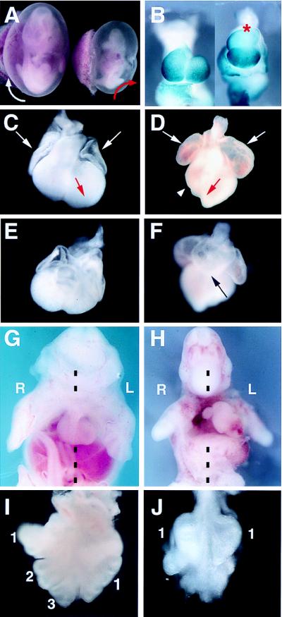
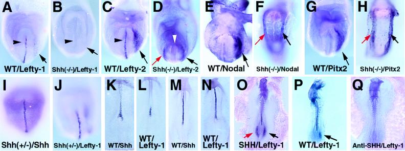
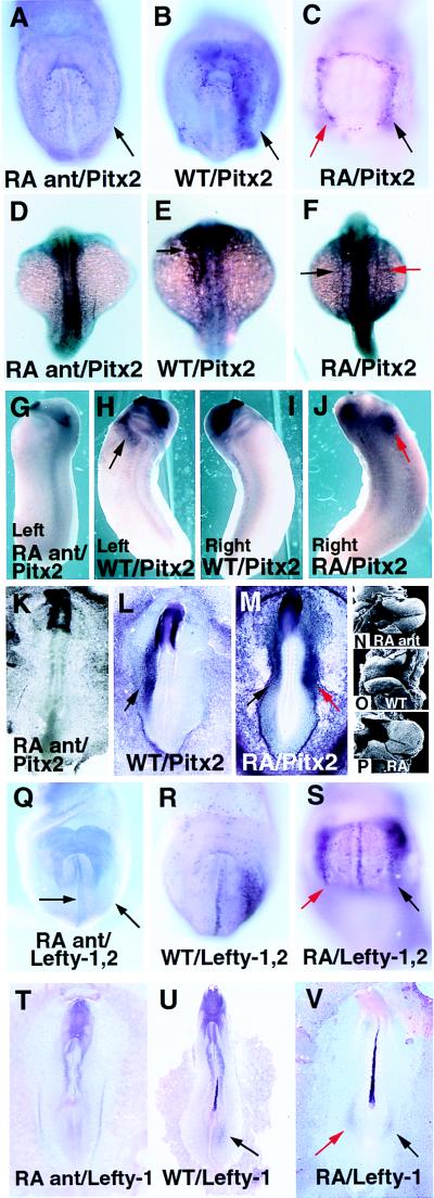
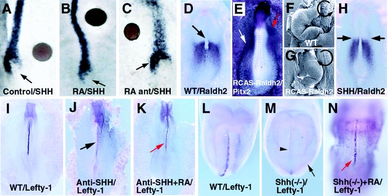
Asymmetric expression of Sonic hedgehog (Shh) in Hensen's node of the chicken embryo plays a key role in the genetic cascade that controls left-right asymmetry, but its involvement in left-right specification in other vertebrates remains unclear. We show that mouse embryos lacking Shh display a variety of laterality defects, including pulmonary left isomerism, alterations of heart looping, and randomization of axial turning. Expression of the left-specific gene Lefty-1 is absent in Shh(-/-) embryos, suggesting that the observed laterality defects could be the result of the lack of Lefty-1. We also demonstrate that retinoic acid (RA) controls Lefty-1 expression in a pathway downstream or parallel to Shh. Further, we provide evidence that RA controls left-right development across vertebrate species. Thus, the roles of Shh and RA in left-right specification indeed are conserved among vertebrates, and the Shh and RA pathways converge in the control of Lefty-1.


Figures

Figure 1
Figure 1
Shh−/− embryos show multiple laterality defects. Shh+/− embryos (C, E, G, and I) are indistinguishable from wild-type ones and from now on will be referred to as such. (A) Shh−/− embryos (Right) display randomization of axial turning (red arrow indicates reversed turning of the tail, which points to the left side of the body in the mutant; compare with the Shh+/− littermate on the left, where the tail points to the right). (B, ventral views) Whole-mount in situ hybridization using a probe to detect the ventricular marker MLC-2V shows a dilated conotruncus (red asterisk) and failure to complete rotation in the Shh−/− heart (compare with the normal heart on the left). (C and D, ventral views) Shh−/− hearts display a severe reduction of the right ventricle (arrowhead in the mutant heart shown in D) and enlarged atriae (arrows in C and D). Shh−/− hearts display an enlarged apex that points toward the right side (red arrow in D, compare with the normal apex pointing toward the left side in the normal heart shown in C). (E and F, dorsal views) Wild-type (E) and Shh−/− (F) heart, where the Shh−/− heart has an abnormally enlarged inflow tract (arrow in F). (G and H) The heart of E12.5 wild-type mice normally is positioned in the midline (G). In Shh mutant mice, the heart is positioned toward the left side of the thoracic cavity (H). R, right; L, left. Broken lines indicate the axes of the embryos. (I and J) In contrast to the wild-type pattern (I), which is four lobes on the right and one on the left (the fourth right lobe is not visible in I), the lungs of Shh−/− mice display a single hypoplastic lobe on each side (J; only the portions marked with 1 are lung tissue).
Figure 2
Figure 2
Shh controls Lefty-1 expression. All embryos are viewed from the ventral side (left side of the embryo corresponds to the reader’s right). Phenotypes or experimental manipulations, followed by the probe used for in situ, are shown at the bottom of each panel, unless otherwise indicated. (A-H) Expression of Lefty-1, Lefty-2, Nodal, and Pitx2 in E8–8.5 Shh−/− embryos. (A and B) Lefty-1 normally is expressed on the left side of the presumptive floor plate (arrowhead in A) and weakly on the left LPM (arrow in A). In Shh−/− mice (B), expression of Lefty-1 is not detected, neither in the midline (arrowhead) nor the left LPM (arrow). (C and D) Lefty-2 also is expressed in the midline (arrowhead) and more strongly on the left LPM (arrow) of wild-type embryos (C). In Shh−/− embryos, Lefty-2 expression in the midline is not detected (white arrowhead), whereas an ectopic induction on the right LPM (red arrow) is observed (D; black arrow shows normal expression in the left LPM). (E and F) Nodal, normally expressed on the left LPM (arrow in E), is ectopically induced on the right LPM of Shh−/− embryos (red arrow in F). (G and H) Similarly, Pitx2, normally expressed on the left LPM (arrow in G) is ectopically induced on the right LPM in Shh−/− embryos (red arrow in H). (I-J) In the mouse, Shh is expressed symmetrically in the head process at E7.5-E8.5 (I). Lefty-1 transcripts are localized on the left side of the perspective floor plate (J). (K and L) In the chick, asymmetric expression of Shh on the left side of the node at stage 5+ coincides with the initial asymmetric expression of Lefty-1. (M and N) In subsequent stages (7 to 9), Lefty-1 and Shh transcripts overlap in a symmetrical pattern in the notochord of the chicken embryo. (O-Q) Shh beads implanted on the right side of the chicken node at stage 6 induce an ectopic patch of Lefty-1 expression that is observed on the posterior right LPM (red arrow in O; black arrow shows normal expression in the posterior left LPM). When beads soaked in a blocking anti-Shh antibody are implanted on the left side of a stage 6 chicken embryo, Lefty-1 expression is neither detected in the midline nor in the posterior left LPM (Q, compare with the normal pattern in P). WT, wild type.
Figure 3
Figure 3
RA alters Lefty and Pitx2 gene expression. (A–C, ventral views) Treatment of mouse embryos for 4–6 hr with the retinoic antagonist AGN 193109 (10−7 M) down-regulates Pitx2 expression on the left LPM (arrow in A; wild type in B). Treatment of mouse embryos with RA (10−7 M) for 4–6 hr at the head fold stage induces Pitx2 expression on the right LPM (red arrow in C). (D–M) Treatment of zebrafish (D–F, dorsal views), Xenopus (G–J, lateral views), and chicken (K–M, dorsal views) with RA antagonist, or RA, down-regulates or induces Pitx2 expression, respectively (black arrows indicate normal expression in the left LPM and red arrows indicate ectopic expression in the right LPM). (N–P) Chicken embryos that were allowed to develop until stage 12 after treatment with RA antagonist (N) or RA (P) display a randomization of heart looping. Here we show hearts looping to the left (compare with the wild-type heart that loops to the right in O). (Q–V, ventral views) Treatment of mouse embryos at the head fold stage with RA antagonist (10−7 M) for 6 hr results in down-regulation of Lefty-1 and Lefty-2 genes in both the midline and LPM (arrows in Q; normal expression in R). On the other hand, similar treatment with RA induces ectopic expression of Lefty genes (red arrow in S). In the chicken embryo, when a bead soaked in RA antagonist is implanted on the left side of the node at stage 6, Lefty-1 expression is abolished (T; normal expression in U). On the contrary, RA beads implanted on the right side of the node of stage 6 chicken embryos induce Lefty-1 expression on the posterior right LPM (red arrow in V; black arrow indicates normal expression on the posterior left LPM). In the dorsal views (D–F and K–M), the left side of the embryo corresponds to the reader’s left). WT, wild type.
Figure 4
Figure 4
Relationship between the Shh and RA pathways. (A-C, dorsal views) Beads soaked in RA (10−7 M, on the right side of the node at stage 4, B), or RA antagonist (10−7 M, on the left side of the node at stage 4, C) do not alter the normal left-sided expression of Shh in the node of the chicken embryos (A; control bead). Beads are the brown circles. (D, dorsal view) Expression pattern of Raldh2 in stage 5+ chicken embryos. Raldh2 transcripts are absent from the node (arrow) and are symmetrically detected on the left and right mesoderm adjacent to the node. (E) Dorsal view. Overexpression of Raldh2 at stage 4, using a retroviral competent vector (RCAS-Raldh2), induces ectopic expression of Pitx2 on the right LPM of chicken embryos (red arrow in E; white arrow indicates normal expression in the left LPM). (F-G) When embryos are allowed to develop until stage 12 after Raldh2 misexpression, we observe a change in the direction of heart looping (in 30% of injected embryos, the heart loops to the left, G; compare with the normal looping to the right in F). Black semicircular arrows indicate direction of heart looping. (H, dorsal view) Application of Shh protein, either to the left or right side of the node at stages 4–5, does not alter the symmetrical expression of Raldh2 (arrows). (I-K, ventral views) Beads soaked in anti-Shh antibody implanted at stage 4 on the left side of the node, down-regulate Lefty-1 expression in the midline (arrow in J, compare with the normal pattern in I). Lefty-1 expression can be rescued (red arrow in K) when a bead soaked in RA is implanted 3 hr after implantation of the anti-Shh bead (compare with normal pattern in I). (L-N, ventral views) Lefty-1 is absent in both the midline (arrowhead) and the left LPM (arrow) of Shh−/− mouse embryos (M, compare with L). When Shh−/− mouse embryos are cultured in the presence of RA (10−7 M for 6 hr at the head fold stage), Lefty-1 expression is restored (red arrow in N). WT, wild type.

References

    1. Harvey R P. Cell. 1998;94:273–276. - PubMed
    1. Levin M, Johnson R L, Stern C D, Kuehn M, Tabin C. Cell. 1995;82:803–814. - PubMed
    1. Ryan A K, Blumberg B, Rodriguez-Esteban C, Yonei-Tamura S, Tamura K, Tsukui T, de la Peña J, Sabbagh W, Greenwald J, Choe S, et al. Nature (London) 1998;394:545–551. - PubMed
    1. Meno C, Shimono A, Saijoh Y, Yashiro K, Mochida K, Ohishi S, Noji S, Kondoh H, Hamada H. Cell. 1998;94:287–297. - PubMed
    1. Pagán-Westphal S M, Tabin C J. Cell. 1998;93:25–35. - PubMed

Publication types

Associated data