. 1999 Sep 28;96(20):11613-8.
doi: 10.1073/pnas.96.20.11613.

IL-1beta differentially regulates calcium wave propagation between primary human fetal astrocytes via pathways involving P2 receptors and gap junction channels

Affiliations

IL-1beta differentially regulates calcium wave propagation between primary human fetal astrocytes via pathways involving P2 receptors and gap junction channels

G R John et al. Proc Natl Acad Sci U S A. .

Abstract

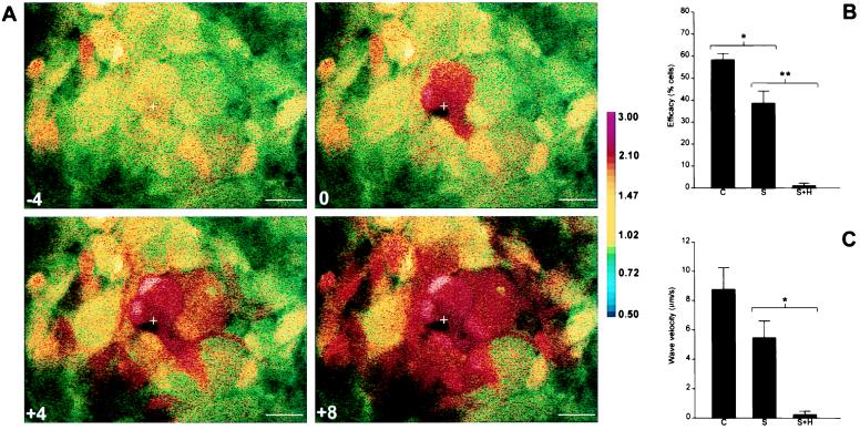
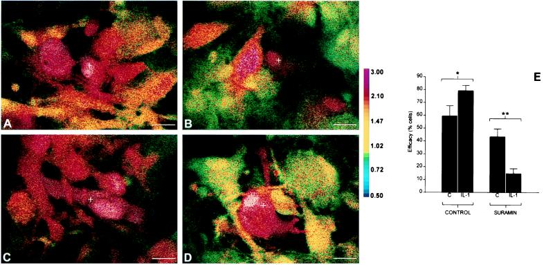
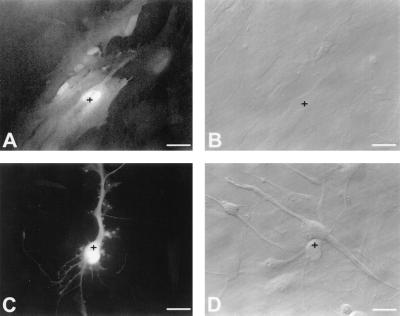
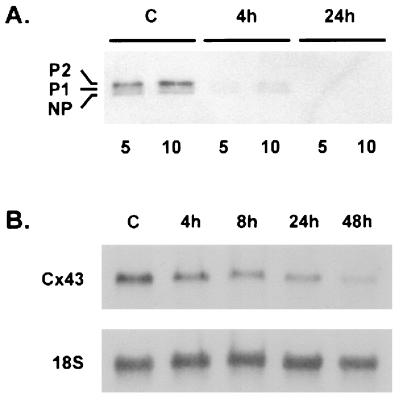
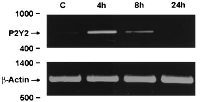
In mammalian astrocytes, calcium waves are transmitted between cells via both a gap junction-mediated pathway and an extracellular, P2 receptor-mediated pathway, which link the cells into a syncytium. Calcium waves in astrocytes have also been shown to evoke calcium transients in neurons, and activity in neurons can elicit calcium waves in astrocytes. In this study, we show that in primary human fetal astrocytes, the P2 receptor-mediated and gap junction-mediated pathways are differentially regulated by the cytokine IL-1beta. Confocal microscopy of astrocytes loaded with Indo-1 demonstrated that intercellular calcium wave transmission in IL-1beta-treated cultures was potentiated compared with controls. However, transmission of calcium waves via the gap junction-mediated pathway was strikingly reduced. The major component of functional gap junctions in human fetal astrocytes was demonstrated to be connexin43 (Cx43), and there was a marked reduction of junctional conductance, loss of dye coupling, loss of Cx43 protein, and down-regulation of Cx43 mRNA expression after IL-1beta treatment of cultures. Conversely, transmission of calcium waves via the P2 receptor-mediated pathway was potentiated in IL-1beta-treated cultures compared with controls. This potentiation was associated with an increase in the number of cells responsive to UTP, and with a transient increase in expression of the P2Y(2) purinoceptor mRNA. Because in inflammatory conditions of the human central nervous system IL-1beta is produced both by resident glia and by invading cells of the immune system, our results suggest that inflammatory events may have a significant impact on coordination of astrocytic function and on information processing in the central nervous system.


Figures

Figure 1
Figure 1
Intercellular calcium wave transmission in cultured human fetal astrocytes. Confluent cultures were loaded with Indo-1AM (A), and a single cell (marked by crosshairs) was mechanically stimulated during ratiometric confocal imaging. The pseudocolor map indicates Indo-1 fluorescence ratio, representative of [Ca2+]i. Time (sec) before and after stimulation is indicated on each panel. (Scale bar = 25 μm.) Data shown are representative of five independent experiments. Efficacy (B) and velocity (C) of wave transmission are shown for representative control cultures, suramin-treated cultures (S), and cultures treated with suramin and heptanol (S+H). Data shown are derived from three separate confluent fields (approximately 15 cells per field) for each condition. ∗, P < 0.05; ∗∗, P < 0.01.
Figure 2
Figure 2
Effect of IL-1β treatment on calcium wave transmission in cultured human fetal astrocytes. Confluent cultures were loaded with Indo-1AM (AD) and subjected to mechanical stimulation as described for Fig. 1. The spread of calcium wave in control cultures in the absence (A) or presence (B) of suramin is shown. The spread in cultures treated with 10 ng/ml IL-1β for 24 hr in the absence (C) or presence (D) of suramin is also shown. Before stimulation, the Indo-1 fluorescence ratio was between 0.8 and 1.0 for all cells in all conditions, and images shown were captured 10 sec after mechanical stimulation, corresponding to maximal extent of wave spread for all conditions. Treatment with IL-1β had striking differential effects on the efficacy of wave transmission in the absence or presence of suramin (E). Note the potentiation of efficacy of wave transmission by IL-1β (∗, P < 0.05), and the striking reduction in wave transmission via the suramin-insensitive pathway (∗∗, P < 0.01). Data shown are derived from six separate confluent fields (approximately 15 cells per field) for each condition and are representative of three independent experiments.
Figure 3
Figure 3
Effect of IL-1β on dye coupling. Individual cells (marked by crosshairs) in confluent control or IL-1β-treated cultures were iontophoretically injected with Lucifer Yellow and photographed 2 min after injection. A and B show matched fluorescence (A) and phase contrast (B) images of injected control cultures, and C and D show matched images from IL-1β-treated cultures. Note the lack of dye transfer from the injected cell in IL-1β-treated cultures. Note also the change in cell morphology induced by IL-1β. Data shown are representative of three independent experiments, at least four cells injected per condition in each experiment. (Scale bar = 25 μm.)
Figure 4
Figure 4
Effect of IL-1β on Cx43 expression. (A) Immunoblot analysis of Cx43 protein expression. Membrane extracts were prepared from control cultures (C) and cultures that had been treated with IL-1β for 4 or 24 hr. Five-and ten-microgram samples were loaded and subjected to SDS/PAGE and immunoblot analysis performed for Cx43. Note that of the three forms (NP, P1, and P2) detected, P2 predominated, and that treatment with IL-1β resulted in loss of all three forms from the membrane. Data are representative of four independent experiments. (B) Northern blot analysis of Cx43 mRNA expression in astrocytes. Total RNA was extracted from untreated cultures (C) and cultures treated with IL-1β for the times indicated, separated by electrophoresis, and hybridized with a 32P-labeled probe specific for Cx43. The blots were then stripped and reprobed for 18S rRNA. Note that treatment with IL-1β led to progressive down-regulation of the Cx43 signal. Data are representative of four independent experiments.
Figure 5
Figure 5
PCR analysis of P2Y2 mRNA expression. Total RNA was extracted from untreated cultures (C) and cultures treated with IL-1β for the times indicated, and subjected to reverse transcription–PCR for P2Y2 and beta-actin as described in the text. Note that treatment with IL-1β led to a transient up-regulation of the signal for the P2Y2 receptor, which was verified by cloning and sequencing. Data are representative of three independent experiments.

References

    1. Sanderson M J, Charles A C, Boitano S, Dirksen E R. Mol Cell Endocrinol. 1994;98:173–187. - PubMed
    1. Saez J C, Connor J A, Spray D C, Bennett M V. Proc Natl Acad Sci USA. 1989;86:2708–2712. - PMC - PubMed
    1. Boitano S, Dirksen E R, Sanderson M J. Science. 1992;258:292–295. - PubMed
    1. Osipchuk Y, Cahalan M. Nature (London) 1992;359:241–244. - PubMed
    1. Yuste R, Nelson D, Rubin W, Katz L C. Neuron. 1995;14:7–17. - PubMed

Publication types