

Simulation model of an eyeball based on finite element analysis on a supercomputer
- PMID: 10502567
- PMCID: PMC1722840
- DOI: 10.1136/bjo.83.10.1106
Simulation model of an eyeball based on finite element analysis on a supercomputer
Abstract
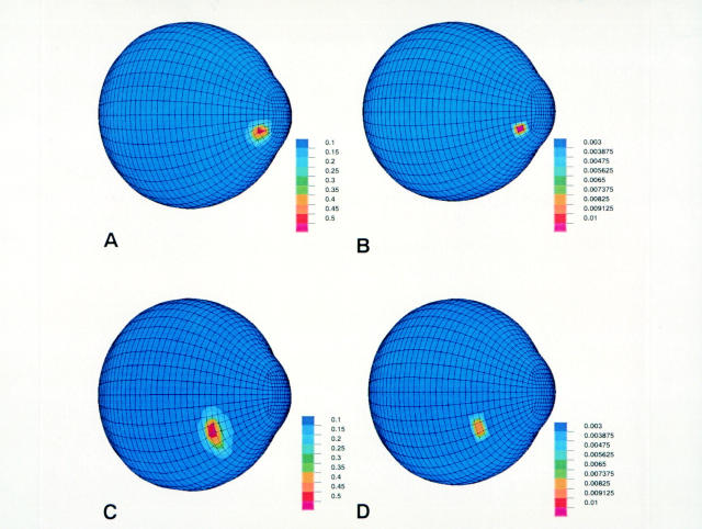
Background/aims: A simulation model of the human eye was developed. It was applied to the determination of the physical and mechanical conditions of impacting foreign bodies causing intraocular foreign body (IOFB) injuries.
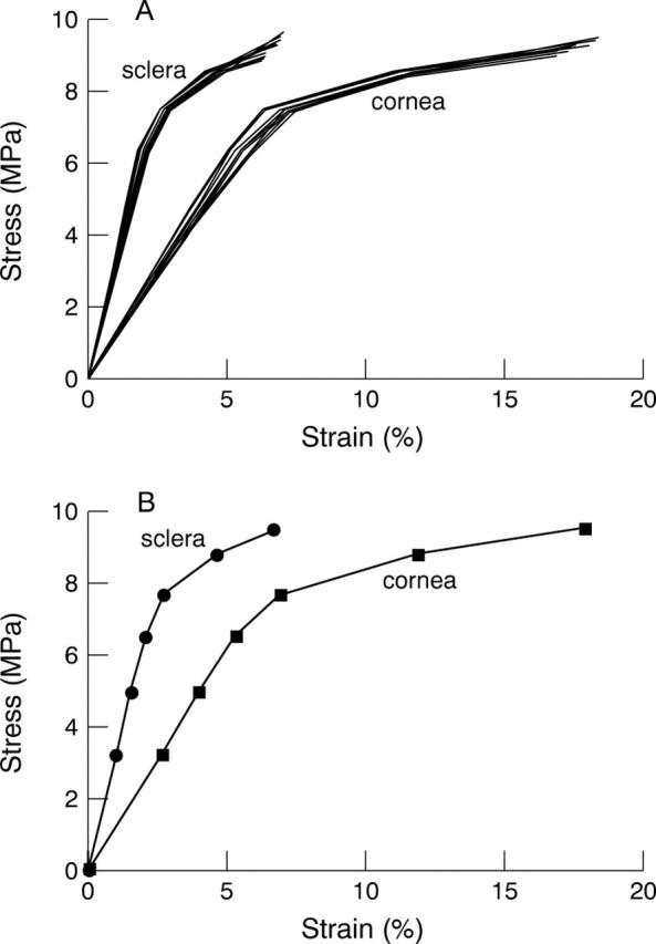
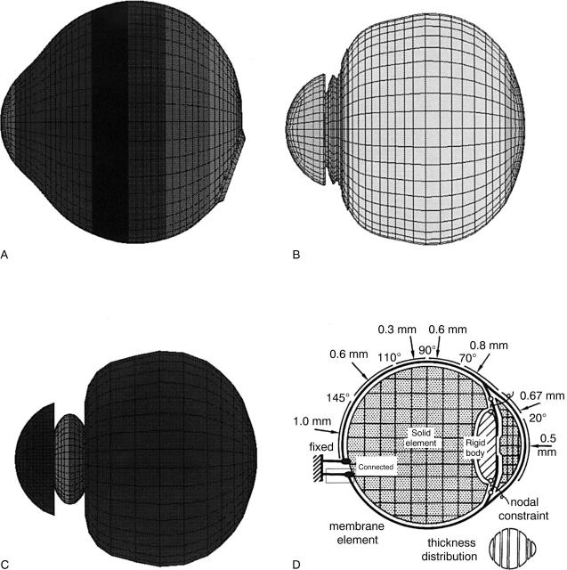
Methods: Modules of the Hypermesh (Altair Engineering, Tokyo, Japan) were used for solid modelling, geometric construction, and finite element mesh creation based on information obtained from cadaver eyes. The simulations were solved by a supercomputer using the finite element analysis (FEA) program PAM-CRASH (Nihon ESI, Tokyo, Japan). It was assumed that rupture occurs at a strain of 18.0% in the cornea and 6.8% in the sclera and at a stress of 9.4 MPa for both cornea and sclera. Blunt-shaped missiles were shot and set to impact on the surface of the cornea or sclera at velocities of 30 and 60 m/s, respectively.
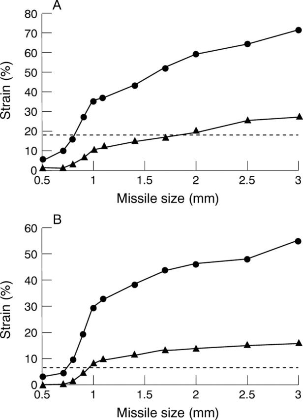
Results: According to the simulation, the sizes of missile above which corneal rupture occurred at velocities of 30 and 60 m/s were 1.95 and 0.82 mm. The missile sizes causing scleral rupture were 0.95 and 0.75 mm at velocities of 30 and 60 m/s.
Conclusions: These results suggest that this FEA model has potential usefulness as a simulation tool for ocular injury and it may provide useful information for developing protective measures against industrial and traffic ocular injuries.
Figures
Comment in
-
Penetrating injury of the eye.Br J Ophthalmol. 1999 Oct;83(10):1101-2. doi: 10.1136/bjo.83.10.1101. Br J Ophthalmol. 1999. PMID: 10502565 Free PMC article. No abstract available.
References
Publication types
MeSH terms
LinkOut - more resources
Full Text Sources
Other Literature Sources
Miscellaneous