

Choroidal folds and papilloedema
- PMID: 10502574
- PMCID: PMC1722841
- DOI: 10.1136/bjo.83.10.1139
Choroidal folds and papilloedema
Abstract
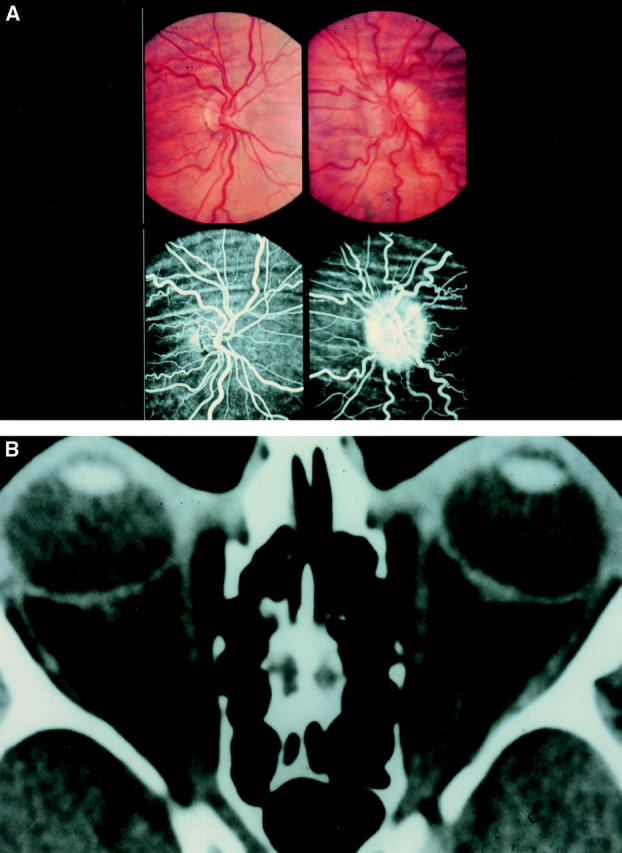
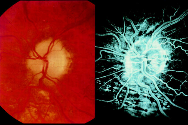
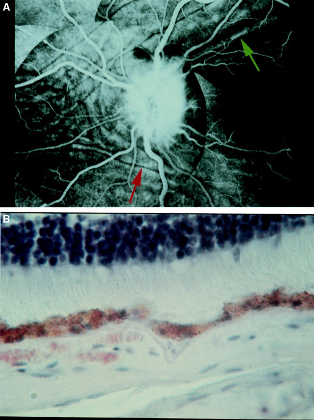
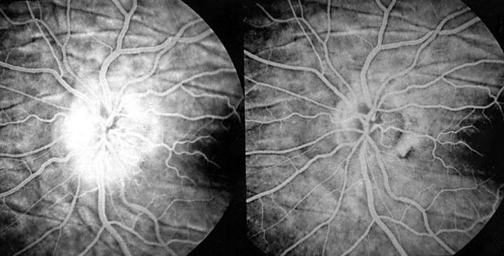
Aims: To assess the clinical and fluorescein angiographic features of choroidal folds seen in association with papilloedema.
Methods: In a retrospective study, the clinical data from a database on patients with choroidal folds (1963-97), including fundus photography and fluorescein angiography, from 32 patients (64 eyes) with choroidal folds in association with papilloedema were reviewed. The clinical and fluorescein angiographic features and the clinical course of choroidal folds in these patients are described.
Results: 32 patients had choroidal folds associated with papilloedema. Folds of two distinct categories were observed, either coarse folds or wrinkles. The folds persisted in all cases, even after resolution of papilloedema. Follow up ranged from 1 month to 20 years. Only one patient suffered permanent visual impairment as a result of a choroidal fold.
Conclusions: Choroidal folds exist in two forms, coarse folds and wrinkles. They persist even after papilloedema has resolved. Final visual acuity did not appear to be affected by the presence of choroidal folds in the majority of patients.
Figures

Similar articles
-
Choroidal folds in association with papilloedema.Br J Ophthalmol. 1973 Feb;57(2):89-97. doi: 10.1136/bjo.57.2.89. Br J Ophthalmol. 1973. PMID: 4735114 Free PMC article. No abstract available.
-
Idiopathic polypoidal choroidal vasculopathy of the macula.Ophthalmology. 1998 Aug;105(8):1380-5. doi: 10.1016/S0161-6420(98)98016-2. Ophthalmology. 1998. PMID: 9709746
-
Intracranial hypertension and the syndrome of acquired hyperopia with choroidal folds.J Neuroophthalmol. 1995 Sep;15(3):178-85. J Neuroophthalmol. 1995. PMID: 8574365
-
[Choroidal folds].Oftalmologia. 2008;52(2):23-8. Oftalmologia. 2008. PMID: 19065910 Review. Romanian.
-
[A new approach for studying the retinal and choroidal circulation].Nippon Ganka Gakkai Zasshi. 2004 Dec;108(12):836-61; discussion 862. Nippon Ganka Gakkai Zasshi. 2004. PMID: 15656089 Review. Japanese.
Cited by
-
The Utility of Fundus Fluorescein Angiography in Neuro-Ophthalmology.Neuroophthalmology. 2019 Aug 21;43(4):217-234. doi: 10.1080/01658107.2019.1604764. eCollection 2019 Aug. Neuroophthalmology. 2019. PMID: 31528186 Free PMC article. Review.
-
Choroidal folds associated with carotid cavernous fistula: a case report.Int J Ophthalmol. 2022 Nov 18;15(11):1881-1884. doi: 10.18240/ijo.2022.11.22. eCollection 2022. Int J Ophthalmol. 2022. PMID: 36404968 Free PMC article. No abstract available.
-
Diagnostic dilemma of papilledema and pseudopapilledema.Int Ophthalmol. 2024 Jun 25;44(1):272. doi: 10.1007/s10792-024-03215-5. Int Ophthalmol. 2024. PMID: 38916684 Review.
-
Orbital fat swelling: A biomechanical theory and supporting model for spaceflight-associated neuro-ocular syndrome (SANS).Front Bioeng Biotechnol. 2023 Feb 9;11:1095948. doi: 10.3389/fbioe.2023.1095948. eCollection 2023. Front Bioeng Biotechnol. 2023. PMID: 36845176 Free PMC article.
-
Retinal and Choroidal Folds in Papilledema.Invest Ophthalmol Vis Sci. 2015 Sep;56(10):5670-80. doi: 10.1167/iovs.15-17459. Invest Ophthalmol Vis Sci. 2015. PMID: 26335066 Free PMC article.
References
MeSH terms
LinkOut - more resources
Full Text Sources