. 1999 Oct;43(10):2457-62.
doi: 10.1128/AAC.43.10.2457.

The new ketolide HMR3647 accumulates in the azurophil granules of human polymorphonuclear cells

Affiliations

The new ketolide HMR3647 accumulates in the azurophil granules of human polymorphonuclear cells

C Miossec-Bartoli et al. Antimicrob Agents Chemother. 1999 Oct.

Abstract

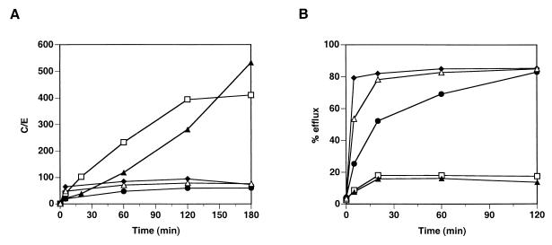
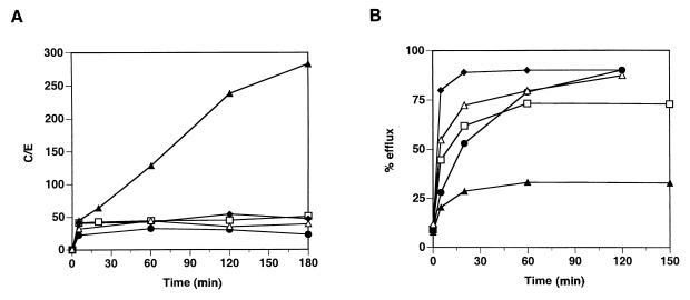
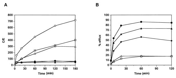
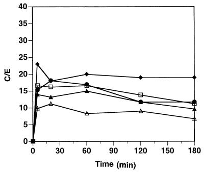
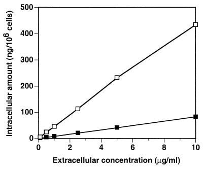
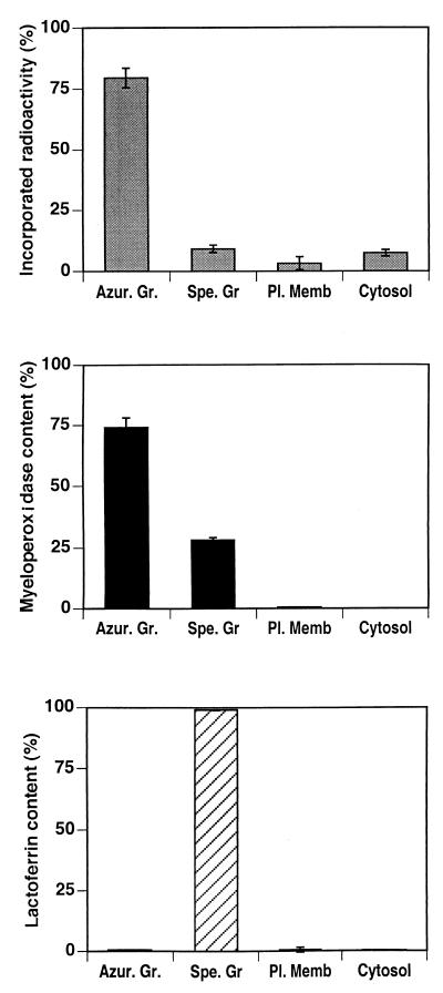
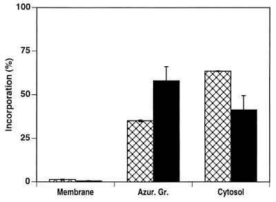
HMR3647 is a semisynthetic representative of a new group of drugs, the ketolides, derived from erythromycin A. Since macrolides have been shown to accumulate in human polymorphonuclear cells (PMNs), we have investigated the ability of the molecule HMR3647 to enter human PMNs as well as other cell types, such as peripheral blood mononuclear cells and cell lines of hematopoietic and nonhematopoietic origin. In these experiments, HMR3647 was compared to erythromycin A, azithromycin, clarithromycin, and roxithromycin. Our results show that HMR3647 is specifically trapped in PMNs, where it is concentrated up to 300 times. In addition, it is poorly released by these cells, 80% of the compound remaining cell associated after 2 h in fresh medium. By contrast, it is poorly internalized and quickly released by the other cell types studied. This differs from the results obtained with the macrolide molecules, which behaved similarly in the different cells studied. In addition, subcellular fractionation of PMNs allowed us to identify the intracellular compartment where HMR3647 was trapped. In PMNs, more than 75% of the molecule was recovered in the azurophil granule fraction. Similarly, in NB4 cells differentiated into PMN-like cells, almost 60% of the molecules accumulated in the azurophil granule fraction. In addition, when HMR3647 was added to disrupted PMNs, 63% accumulated in the azurophil granules. Therefore, this study shows that the ketolide HMR3647 specifically accumulates in PMN azurophil granules, thus favoring its delivery to bacteria phagocytosed in these cells.


Figures

FIG. 1
FIG. 1
Incorporation and release of HMR3647 and macrolide comparators into PMNs. Human PMNs were incubated with radiolabelled HMR3647 (open squares), azithromycin (solid triangles), clarithromycin (solid diamonds), erythromycin (solid circles), and roxithromycin (open triangles) at 10-μg/ml final concentration. (A) Drug uptake was monitored at different time points and is expressed as a C/E ratio. (B) Drug release from the cells was monitored after 2 h of incubation with the radiolabelled compounds and is expressed as the percentage of drug found in the supernatant (% efflux). The results correspond to the mean of three independent experimental points obtained with cells from the same blood donor. Standard errors of the mean were always below 10%.
FIG. 2
FIG. 2
Incorporation and release of HMR3647 and macrolide comparators into PBMC. Human PBMC were incubated with radiolabelled HMR3647 (open squares), azithromycin (solid triangles), clarithromycin (solid diamonds), erythromycin (solid circles), and roxithromycin (open triangles) at 10-μg/ml final concentration. (A) Drug uptake was monitored as described in the legend to Fig. 1. The results correspond to the mean of three independent experimental points obtained with cells from the same blood donor. Standard errors of the mean were always below 10%.
FIG. 3
FIG. 3
Incorporation and release of HMR3647 in PMNs and PBMC from different individuals. Human PMNs (open symbols) and PBMC (solid symbols) were obtained from different blood donors. HMR3647 uptake (A) and release (B) were monitored as described in the legend to Fig. 1. Each datum point corresponds to the mean of three independent experimental points obtained with cells from the same blood donor. Standard errors of the mean were always below 10%.
FIG. 4
FIG. 4
Incorporation of HMR3647 into human cell lines. Jurkat (solid squares), K562 (solid triangles), THP-1 (open circles), NB4 (solid diamonds), and Colo205 (open squares) cells were incubated with radiolabelled HMR3647 (10-μg/ml final concentration). Drug uptake by the cells was monitored at different time points and is expressed as the C/E ratio. The results correspond to the means of three independent experimental points obtained with cells from the same blood donor. Standard errors of the mean were always below 10%.
FIG. 5
FIG. 5
Incorporation of HMR3647 in PMNs: effect of extracellular drug concentration. Human PMNs were incubated for 5 min (solid squares) or 120 min (open squares) with radiolabelled HMR3647 at different concentrations (0.1, 1, 2.5, 5, and 10 μg/ml). Drug incorporation into the cells is expressed as the amount of drug (in nanograms) present in 106 cells. The results correspond to the means of three independent experimental points obtained with cells from the same blood donor. Standard errors of the mean were always below 10%.
FIG. 6
FIG. 6
Accumulation of HMR3647 in PMN azurophil granules. Human PMNs were incubated for 120 min with radiolabelled HMR3647 (10-μg/ml final concentration) and then disrupted by nitrogen cavitation and fractionated on a discontinuous Percoll gradient. The radioactivity incorporated into the membrane fractions (Azur. Gr, azurophil granules; Spe. Gr, specific granules; Pl. Memb, plasma membrane) and the cytosol was counted. Markers for azurophil granules (myeloperoxidase) and specific granules (lactoferrin) were assayed in each fraction by enzyme-linked immunosorbent assay. The data are expressed as mean ± standard error of the mean of four experiments.
FIG. 7
FIG. 7
HMR3647 accumulates in azurophil granules only in NB4 cells differentiated into PMN-like cells. NB4 cells were differentiated for 5 days in the presence of all-trans retinoic acid. Differentiated (solid bars) and nondifferentiated (crosshatched bars) cells were exposed to HMR3647 as described in the legend to Fig. 6 and fractionated by differential centrifugation. The radioactivity incorporated into the different fractions was counted, and the results are expressed as percentages of the radioactivity present in the postnuclear supernatant. The data are expressed as mean ± standard error of the mean of two experiments.

References

    1. Agouridas C, Benedetti Y, Denis A, Le Martret O, Chantot J-F. Program and abstracts of the 35th Interscience Conference on Antimicrobial Agents and Chemotherapy. Washington, D.C: American Society for Microbiology; 1995. Ketolides: a new distinct class of macrolide antibacterials. Synthesis and structural characteristics of RU 004, abstr. 157; p. 140.
    1. Barry A L, Brown S D, Fuchs P C. Program and abstracts of the 37th Interscience Conference on Antimicrobial Agents and Chemotherapy. Washington, D.C: American Society for Microbiology; 1997. Antipneumococcal activity of a ketolide (HMR3647) and seven related drugs in vivo, abstr. 107; p. 164.
    1. Bonnet M, Van der Auwera P. In vitro and in vivo intraleukocytic accumulation of azithromycin (CP-62,993) and its influence on ex vivo leukocyte chemiluminescence. Antimicrob Agents Chemother. 1992;36:1302–1309. - PMC - PubMed
    1. Borregaard N. Current concepts about neutrophil granule physiology. Curr Opin Hematol. 1996;3:11–18. - PubMed
    1. Borregaard N, Cowland J B. Granules of the human neutrophilic polymorphonuclear leukocyte. Blood. 1997;89:3503–3521. - PubMed