

Toxigenic strains of Bacillus licheniformis related to food poisoning
- PMID: 10508100
- PMCID: PMC91618
- DOI: 10.1128/AEM.65.10.4637-4645.1999
Toxigenic strains of Bacillus licheniformis related to food poisoning
Abstract
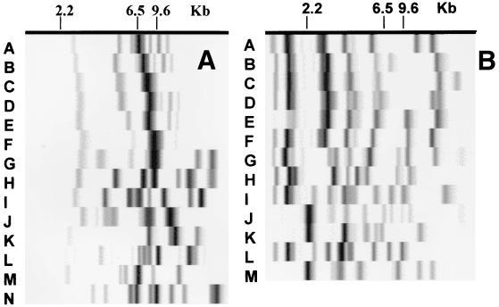
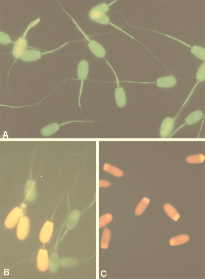
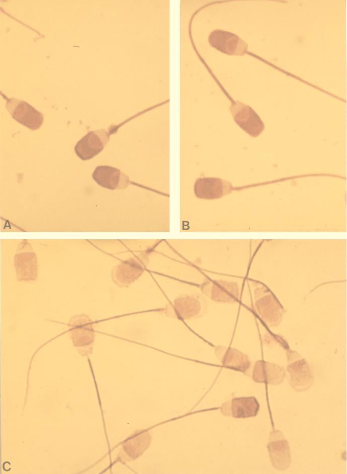
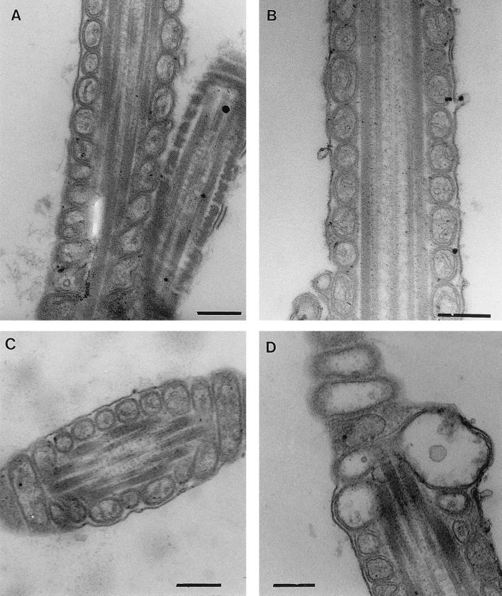
Toxin-producing isolates of Bacillus licheniformis were obtained from foods involved in food poisoning incidents, from raw milk, and from industrially produced baby food. The toxin detection method, based on the inhibition of boar spermatozoan motility, has been shown previously to be a sensitive assay for the emetic toxin of Bacillus cereus, cereulide. Cell extracts of the toxigenic B. licheniformis isolates inhibited sperm motility, damaged cell membrane integrity, depleted cellular ATP, and swelled the acrosome, but no mitochondrial damage was observed. The responsible agent from the B. licheniformis isolates was partially purified. It showed physicochemical properties similar to those of cereulide, despite having very different biological activity. The toxic agent was nonproteinaceous; soluble in 50 and 100% methanol; and insensitive to heat, protease, and acid or alkali and of a molecular mass smaller than 10,000 g mol(-1). The toxic B. licheniformis isolates inhibited growth of Corynebacterium renale DSM 20688(T), but not all inhibitory isolates were sperm toxic. The food poisoning-related isolates were beta-hemolytic, grew anaerobically and at 55 degrees C but not at 10 degrees C, and were nondistinguishable from the type strain of B. licheniformis, DSM 13(T), by a broad spectrum of biochemical tests. Ribotyping revealed more diversity; the toxin producers were divided among four ribotypes when cut with PvuII and among six when cut with EcoRI, but many of the ribotypes also contained nontoxigenic isolates. When ribotyped with PvuII, most toxin-producing isolates shared bands at 2.8 +/- 0.2, 4.9 +/- 0.3, and 11.7 +/- 0.5 or 13.1 +/- 0.8 kb.
Figures
Similar articles
-
Toxic Bacillus pumilus from indoor air, recycled paper pulp, Norway spruce, food poisoning outbreaks and clinical samples.Syst Appl Microbiol. 2001 Jul;24(2):267-76. doi: 10.1078/0723-2020-00025. Syst Appl Microbiol. 2001. PMID: 11518331
-
Toxinogenic Bacillus pumilus and Bacillus licheniformis from mastitic milk.Vet Microbiol. 2007 Oct 6;124(3-4):329-39. doi: 10.1016/j.vetmic.2007.05.015. Epub 2007 May 24. Vet Microbiol. 2007. PMID: 17611049
-
In vitro assay for human toxicity of cereulide, the emetic mitochondrial toxin produced by food poisoning Bacillus cereus.Toxicol In Vitro. 2003 Oct-Dec;17(5-6):737-44. doi: 10.1016/s0887-2333(03)00096-1. Toxicol In Vitro. 2003. PMID: 14599471
-
Bacillus and relatives in foodborne illness.J Appl Microbiol. 2012 Mar;112(3):417-29. doi: 10.1111/j.1365-2672.2011.05204.x. Epub 2011 Dec 20. J Appl Microbiol. 2012. PMID: 22121830 Review.
-
Properties of Bacillus cereus and other bacilli contaminating biomaterial-based industrial processes.Int J Food Microbiol. 2000 Sep 25;60(2-3):231-9. doi: 10.1016/s0168-1605(00)00313-5. Int J Food Microbiol. 2000. PMID: 11016612 Review.
Cited by
-
Toxin-producing ability among Bacillus spp. outside the Bacillus cereus group.Appl Environ Microbiol. 2005 Mar;71(3):1178-83. doi: 10.1128/AEM.71.3.1178-1183.2005. Appl Environ Microbiol. 2005. PMID: 15746316 Free PMC article.
-
Microbiological Food Safety of Seaweeds.Foods. 2021 Nov 6;10(11):2719. doi: 10.3390/foods10112719. Foods. 2021. PMID: 34829000 Free PMC article. Review.
-
The Prevalence and Control of Bacillus and Related Spore-Forming Bacteria in the Dairy Industry.Front Microbiol. 2015 Dec 21;6:1418. doi: 10.3389/fmicb.2015.01418. eCollection 2015. Front Microbiol. 2015. PMID: 26733963 Free PMC article. Review.
-
γ-PGA-Rich Chungkookjang, Short-Term Fermented Soybeans: Prevents Memory Impairment by Modulating Brain Insulin Sensitivity, Neuro-Inflammation, and the Gut-Microbiome-Brain Axis.Foods. 2021 Jan 21;10(2):221. doi: 10.3390/foods10020221. Foods. 2021. PMID: 33494481 Free PMC article. Review.
-
Sequential Separation of Essential Oil Components during Hydrodistillation of Fresh Foliage from Azorean Cryptomeria japonica (Cupressaceae): Effects on Antibacterial, Antifungal, and Free Radical Scavenging Activities.Plants (Basel). 2024 Jun 22;13(13):1729. doi: 10.3390/plants13131729. Plants (Basel). 2024. PMID: 38999569 Free PMC article.
References
-
- Agata N, Ohta M, Masashi M, Isobe M. A novel dodecadepsipeptide, cereulide, is an emetic toxin of Bacillus cereus. FEMS Microbiol Lett. 1995;129:17–20. - PubMed
-
- Andersson M, Laukkanen M, Nurmiaho-Lassila E-L, Rainey F A, Niemelä S I, Salkinoja-Salonen M S. Bacillus thermosphaericus, sp. nov., a new thermophilic ureolytic Bacillus isolated from air. Syst Appl Microbiol. 1995;18:203–220.
-
- de Boer S A, Priest F G, Diedrichsen B. On the industrial use of Bacillus licheniformis: a review. Appl Microbiol Biotechnol. 1994;40:595–598.
Publication types
MeSH terms
LinkOut - more resources
Full Text Sources
Medical
Molecular Biology Databases
