. 1999 Oct 4;147(1):7-12.
doi: 10.1083/jcb.147.1.7.

ADP-ribosylation factor 6 and endocytosis at the apical surface of Madin-Darby canine kidney cells

Affiliations

ADP-ribosylation factor 6 and endocytosis at the apical surface of Madin-Darby canine kidney cells

Y Altschuler et al. J Cell Biol. .

Abstract

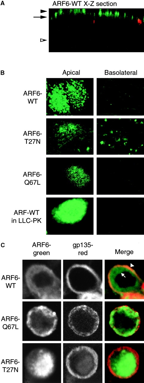
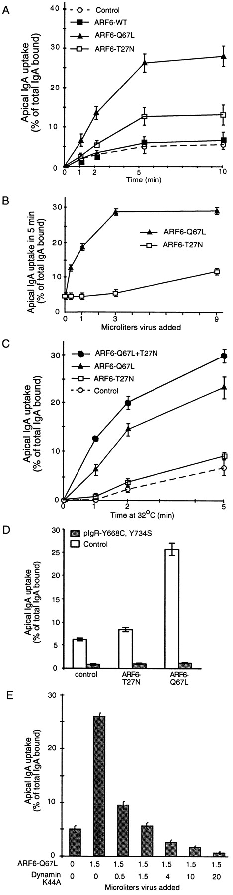
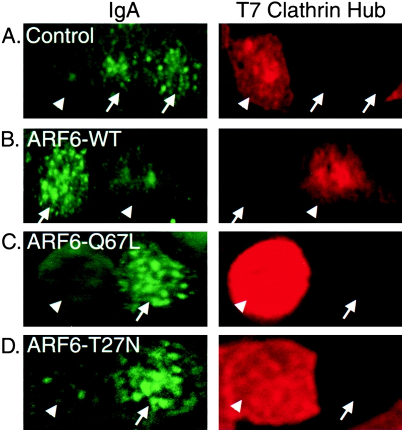
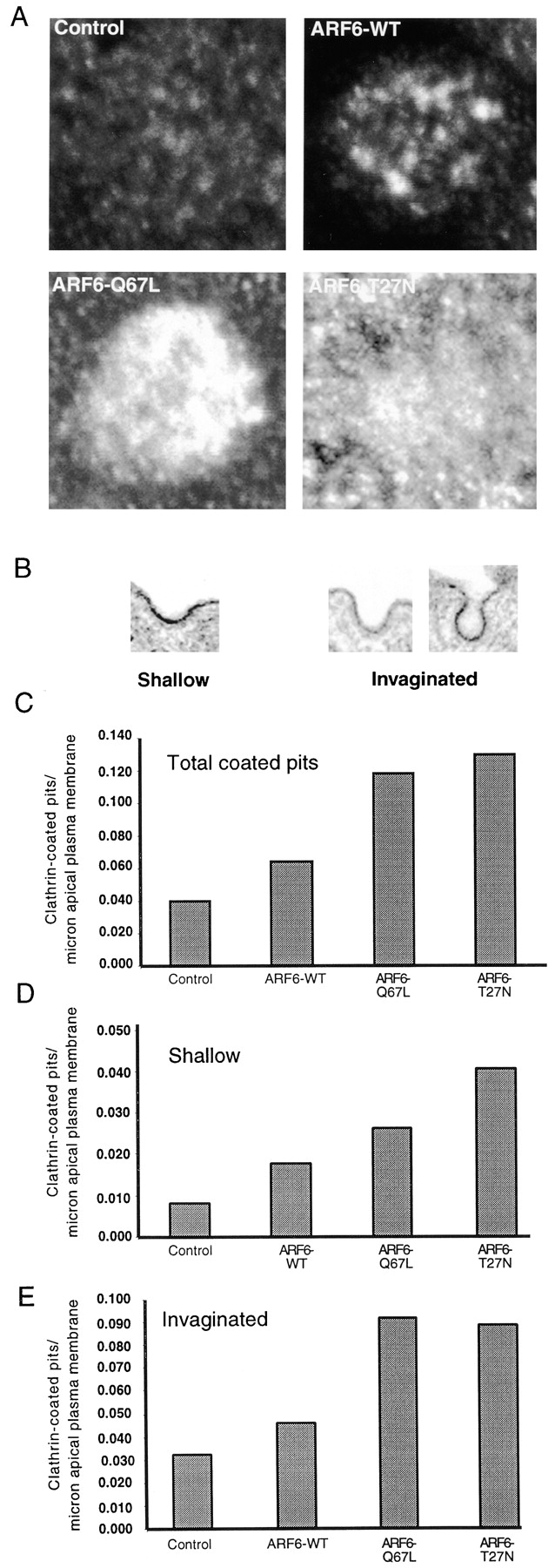
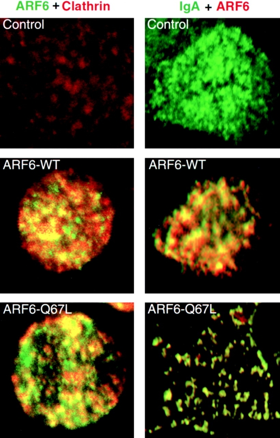
We report that the small GTPase, ADP-ribosylation factor 6 (ARF6), is present only on the apical surface of polarized MDCK epithelial cells. Overexpression of a mutant of ARF6, ARF6-Q67L, which is predicted to be in the GTP-bound form, stimulates endocytosis exclusively at this surface. Surprisingly, overexpression of the mutant ARF6-T27N, which is predicted to be in the GDP-bound form, also stimulated apical endocytosis, though to a lesser extent. ARF6-stimulated endocytosis is inhibited by a dominant-negative form of dynamin, or a dominant-negative hub fragment of clathrin heavy chain, indicating that it is mediated by clathrin. Correspondingly, overexpression of either mutant of ARF6 leads to an increase in the number of clathrin-coated pits at the apical plasma membrane. When ARF6-Q67L is overexpressed in the presence of the dominant-negative dynamin, the ARF6-Q67L colocalizes with clathrin and with IgA bound to its receptor. We conclude that ARF6 is an important modulator of clathrin-mediated endocytosis at the apical surface of epithelial cells.


Figures

Figure 1
Figure 1
Localization of ARF6 in MDCK cells. A, X-Z section of ARF6–WT: ARF6, green (12CA5 antibody to HA epitope); Z0-1, red. B, X-Y sections taken at AP PM (level of solid arrowhead in A) or BL region (level of open arrowhead in A) of cells expressing indicated protein. ARF6–WT was also expressed in LLCPK1 cells, indicating that the AP localization of ARF6–WT is not unique to MDCK cells. C, X-Y sections taken below apical PM, at level of arrow in A.
Figure 2
Figure 2
Effect of ARF6 on AP endocytosis. A, Effect on rate of endocytosis. B, Effects of varying expression level of ARF6 mutants on AP endocytosis. Cells were infected with the indicated quantity of virus. Endocytosis was assayed for 5 min at 37°C, 1 μl of virus ∼25 pfu/cell. C, Effect of coexpression of ARF6 mutants. Cells were either infected with virus for ARF6–Q67L plus virus for β-gal (to keep the total virus constant), ARF6–T27N plus β-gal virus, or doubly infected with viruses encoding both ARF6 mutants. To improve temporal resolution, this experiment was conducted at 32°C. D, Effect of ARF6 on endocytosis of endocytosis-deficient mutant pIgR. Endocytosis of pIgR with both cytoplasmic Tyr mutated to Ala was assayed in control cells or cells expressing ARF6–WT or mutants. In this experiment only, normal MDCK cells stably expressing the pIgR-664A, 734A, or wild-type pIgR were coinfected with the virus for β-gal or ARF6 mutant, plus 70–80 pfu/cell of adenovirus encoding the transactivator. The level of expression of the ARF6 mutant proteins was comparable to other experiments. E, Cells were doubly infected with ARF6–Q67L and the indicated quantity of virus for dynamin-I K44A. Endocytosis over 5 min was measured. Expression of dynamin did not alter expression of the ARF6, as assayed by immunoblot (not shown).
Figure 3
Figure 3
Clathrin hub inhibits ARF6 stimulated AP endocytosis. Cells were infected with 150 pfu/cell of virus expressing the dominant-negative clathrin hub alone, or also with virus for ARF6–WT or mutants. Cells were incubated with IgA at the AP PM for 60 min at 4°C, then at 37°C for 5 min. IgA remaining on the PM was stripped with trypsin at 4°C, under conditions that remove >99% of PM-bound IgA. After fixation and permeabilization, IgA was stained with antibodies to human IgA, while the clathrin hub was stained with an antibody to its T7 epitope tag. Arrowheads indicate cells expressing a high level of hub, which do not endocytose IgA. Arrows indicate cells in which clathrin hub was not detected; these cells do endocytose IgA. All cells expressed pIgR on their AP PM.
Figure 4
Figure 4
ARF6 recruits clathrin to the AP PM. A, Cells were either uninfected or infected with adenovirus for ARF6–WT or mutants, then stained for clathrin. Confocal sections are at the level of the arrowhead in Fig. 1 A. B, Examples of shallow and invaginated clathrin-coated pits, as seen by EM. C, Total coated pits/μm AP PM. D, Shallow coated pits. E, Invaginated coated pits.
Figure 5
Figure 5
Colocalization of ARF6, clathrin, and IgA in cells expressing dynamin-I K44A. Left, Cells were infected with viruses for dynamin-I K44A (100 pfu/cell), and either ARF6–WT or ARF6–Q67L. Cells were treated for 5 min at 4°C with 120 μg/ml digitonin 10 to extract cytoplasmic clathrin, washed three times with cold PBS, fixed, and processed for immunofluorescence to detect ARF6 and endogenous clathrin. Right, Cells were infected with viruses for pIgR, dynamin-I K44A, and either β-gal, ARF6–WT, or ARF6–Q67L. IgA was bound to the AP PM for 1 h at 4°C. Cells were warmed to 15°C for 10 min. Unpermeabilized cells were stained for IgA, then cells were extracted with digitonin, fixed, permeabilized, and ARF6 stained.

References

    1. Altschuler Y., Barbas S.M., Terlecky L.J., Tang K., Hardy S., Mostov K.E., Schmid S.L. Redundant and distinct functions for dynamin-1 and dynamin-2 isoforms. J. Cell Biol. 1998;143:1871–1881. - PMC - PubMed
    1. Apodaca G., Aroeti B., Tang K., Mostov K.E. Brefeldin-A inhibits the delivery of the polymeric immunoglobulin receptor to the basolateral surface of MDCK cells. J. Biol. Chem. 1993;268:20380–20385. - PubMed
    1. Boman A.L., Kahn R.A. Arf proteinsthe membrane traffic police? Trends Biochem. Sci. 1995;20:147–150. - PubMed
    1. Breitfeld P.P., Harris J.M., Mostov K.M. Postendocytotic sorting of the ligand for the polymeric immunoglobulin receptor in Madin-Darby canine kidney cells. J. Cell Biol. 1989;109:475–486. - PMC - PubMed
    1. Cavenaugh M.M., Whitney J.A., Carroll K., Zhang C.-J., Boman A.L., Rosenwald A.G., Mellman I., Kahn R.A. Intracellular distribution of Arf proteins in mammalian cells. J. Biol. Chem. 1996;271:21767–21774. - PubMed

Publication types

MeSH terms