

ErbB4 signaling in the mammary gland is required for lobuloalveolar development and Stat5 activation during lactation
- PMID: 10508857
- PMCID: PMC2164978
- DOI: 10.1083/jcb.147.1.77
ErbB4 signaling in the mammary gland is required for lobuloalveolar development and Stat5 activation during lactation
Abstract
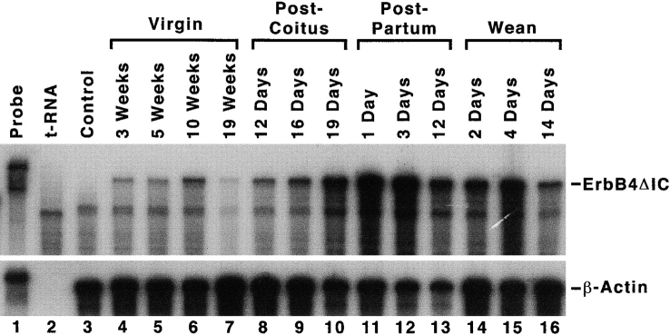
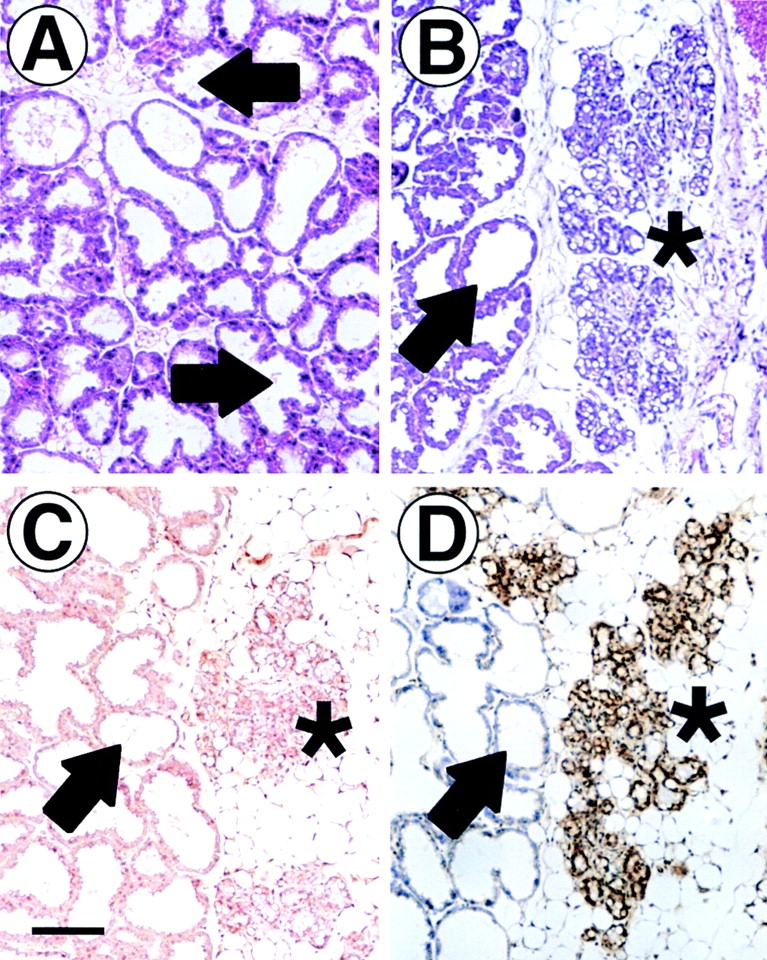
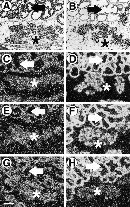
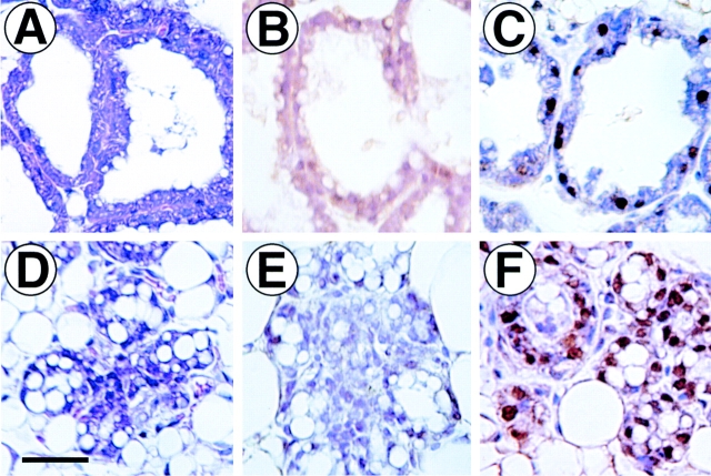
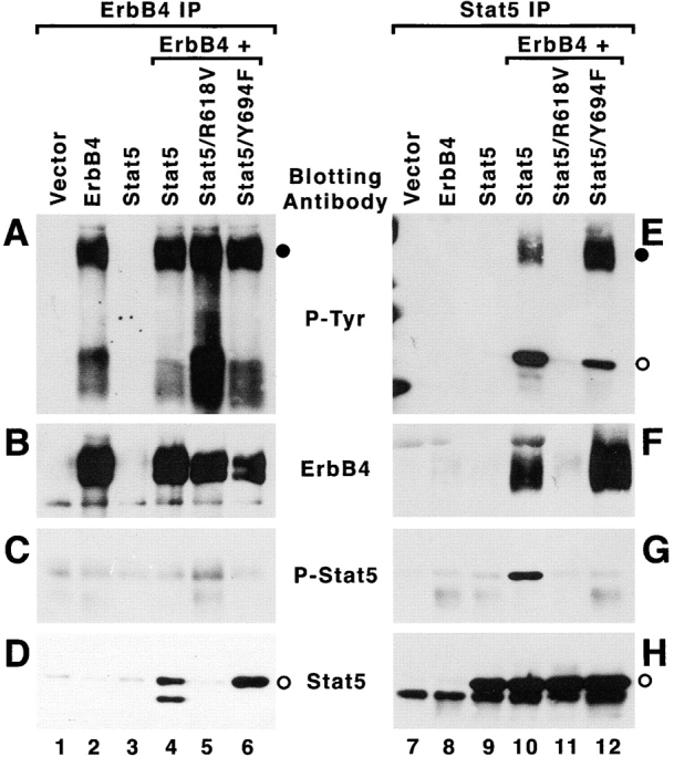
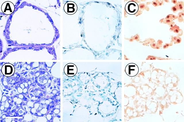
Signaling by members of the epidermal growth factor receptor family plays an important role in breast development and breast cancer. Earlier work suggested that one of these receptors, ErbB4, is coupled to unique responses in this tissue. To determine the function of ErbB4 signaling in the normal mouse mammary gland, we inactivated ErbB4 signaling by expressing a COOH terminally deleted dominant-negative allele of ErbB4 (ErbB4DeltaIC) as a transgene in the mammary gland. Despite the expression of ErbB4DeltaIC from puberty through later stages of mammary development, an ErbB4DeltaIC-specific phenotype was not observed until mid-lactation. At 12-d postpartum, lobuloalveoli expressing ErbB4DeltaIC protein were condensed and lacked normal lumenal lactation products. In these lobuloalveoli, beta-casein mRNA, detected by in situ hybridization, was normal. However, whey acidic protein mRNA was reduced, and alpha-lactalbumin mRNA was undetectable. Stat5 expression was detected by immunohistochemistry in ErbB4DeltaIC-expressing tissue. However, Stat5 was not phosphorylated at Y694 and was, therefore, probably inactive. When expressed transiently in 293T cells, ErbB4 induced phosphorylation of Stat5. This phosphorylation required an intact Stat5 SH2 domain. In summary, our results demonstrate that ErbB4 signaling is necessary for mammary terminal differentiation and Stat5 activation at mid-lactation.
Figures
Similar articles
-
Impaired differentiation and lactational failure of Erbb4-deficient mammary glands identify ERBB4 as an obligate mediator of STAT5.Development. 2003 Nov;130(21):5257-68. doi: 10.1242/dev.00715. Epub 2003 Sep 3. Development. 2003. PMID: 12954715
-
Overexpression and forced activation of stat5 in mammary gland of transgenic mice promotes cellular proliferation, enhances differentiation, and delays postlactational apoptosis.Mol Cancer Res. 2002 Nov;1(1):32-47. Mol Cancer Res. 2002. PMID: 12496367
-
Essential functions of p21-activated kinase 1 in morphogenesis and differentiation of mammary glands.J Cell Biol. 2003 May 12;161(3):583-92. doi: 10.1083/jcb.200212066. Epub 2003 May 5. J Cell Biol. 2003. PMID: 12732616 Free PMC article.
-
Composite response elements mediate hormonal and developmental regulation of milk protein gene expression.Biochem Soc Symp. 1998;63:101-13. Biochem Soc Symp. 1998. PMID: 9513715 Review.
-
ErbB4/HER4: role in mammary gland development, differentiation and growth inhibition.J Mammary Gland Biol Neoplasia. 2008 Jun;13(2):235-46. doi: 10.1007/s10911-008-9080-x. Epub 2008 Apr 25. J Mammary Gland Biol Neoplasia. 2008. PMID: 18437540 Free PMC article. Review.
Cited by
-
Epithelial Xbp1 is required for cellular proliferation and differentiation during mammary gland development.Mol Cell Biol. 2015 May;35(9):1543-56. doi: 10.1128/MCB.00136-15. Epub 2015 Feb 23. Mol Cell Biol. 2015. PMID: 25713103 Free PMC article.
-
Embryonic lethality and defective mammary gland development of activator-function impaired conditional knock-in Erbb3 V943R mice.Adv Genet (Hoboken). 2020 Dec 5;2(1):e10036. doi: 10.1002/ggn2.10036. eCollection 2021 Mar. Adv Genet (Hoboken). 2020. PMID: 36618440 Free PMC article.
-
Impaired alveologenesis and maintenance of secretory mammary epithelial cells in Jak2 conditional knockout mice.Mol Cell Biol. 2004 Jun;24(12):5510-20. doi: 10.1128/MCB.24.12.5510-5520.2004. Mol Cell Biol. 2004. PMID: 15169911 Free PMC article.
-
In vivo evidence for epidermal growth factor receptor (EGFR)-mediated release of prolactin from the pituitary gland.J Biol Chem. 2011 Nov 11;286(45):39297-306. doi: 10.1074/jbc.M111.243493. Epub 2011 Sep 13. J Biol Chem. 2011. PMID: 21914800 Free PMC article.
-
Neuregulin 1-activated ERBB4 interacts with YAP to induce Hippo pathway target genes and promote cell migration.Sci Signal. 2014 Dec 9;7(355):ra116. doi: 10.1126/scisignal.2005770. Sci Signal. 2014. PMID: 25492965 Free PMC article.
References
-
- Alroy I., Yarden Y. The ErbB signaling network in embryogenesis and oncogenesissignal diversification through combinatorial ligand-receptor interactions. FEBS Lett. 1997;410:83–86. - PubMed
-
- Bacus S.S., Zelnick C.R., Plowman G., Yarden Y. Expression of the erbB-2 family of growth factor receptors and their ligands in breast cancersimplication for tumor biology and clinical behavior. Am. J. Clin. Pathol. 1994;102:S13–S24. - PubMed
-
- Berlanga J.J., Garcia-Ruiz J.P., Perrot-Applanat M., Kelly P.A., Edery M. The short form of the prolactin (PRL) receptor silences PRL induction of the β-casein gene promoter. Mol. Endocrinol. 1997;11:1449–1457. - PubMed
-
- Bobrow L.G., Millis R.R., Happerfield L.C., Gullick W.J. c-erbB-3 protein expression in ductal carcinoma in situ of the breast. Eur. J. Cancer. 1997;33:1846–1850. - PubMed
Publication types
MeSH terms
Substances
Grants and funding
LinkOut - more resources
Full Text Sources
Other Literature Sources
Molecular Biology Databases
Research Materials
Miscellaneous
