. 1999 Oct 4;147(1):175-84.
doi: 10.1083/jcb.147.1.175.

Plasma membrane microdomains act as concentration platforms to facilitate intoxication by aerolysin

Affiliations

Plasma membrane microdomains act as concentration platforms to facilitate intoxication by aerolysin

L Abrami et al. J Cell Biol. .

Abstract

It has been proposed that the plasma membrane of many cell types contains cholesterol-sphingolipid-rich microdomains. Here, we analyze the role of these microdomains in promoting oligomerization of the bacterial pore-forming toxin aerolysin. Aerolysin binds to cells, via glycosyl phosphatidylinositol-anchored receptors, as a hydrophilic soluble protein that must polymerize into an amphipathic ring-like complex to form a pore. We first show that oligomerization can occur at >10(5)-fold lower toxin concentration at the surface of living cells than in solution. Our observations indicate that it is not merely the number of receptors on the target cell that is important for toxin sensitivity, but their ability to associate transiently with detergent resistant microdomains. Oligomerization appears to be promoted by the fact that the toxin bound to its glycosyl phosphatidylinositol-anchored receptors, can be recruited into these microdomains, which act as concentration devices.


Figures
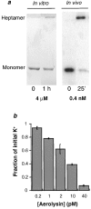
Figure 1
Figure 1
Oligomerization of aerolysin is more efficient in vivo than in vitro. a, To follow oligomerization in vitro, proaerolysin at a concentration of 4 μM was activated with trypsin as described in Materials and Methods. The sample was then incubated at 37°C for 1 h and analyzed by SDS-PAGE, followed by Coomassie staining. The aerolysin heptamer (333 kD) is SDS resistant and can be visualized at the top of the gel. For the in vivo oligomerization experiment, 125I-proaerolysin at a concentration of 0.4 nM was activated in solution with trypsin and added to BHK cells at 4°C for 1 h. Cells were then washed and incubated for 25 min at 37°C. A PNS was prepared and analyzed by SDS-PAGE, followed by autoradiography. b, To measure potassium efflux from BHK cells at different toxin concentrations, cells were incubated with aerolysin for 1 h at 37°C and the cellular potassium contents were determined by flame photometry. Experiments were done in triplicate and the SDs were calculated.
Figure 2
Figure 2
Proaerolysin, aerolysin, and heptameric aerolysin are enriched in Triton X-100 insoluble microdomains. 125I-proaerolysin or trypsin-activated 125I-aerolysin (0.4 nM) were bound to cells at 4°C for 1 h. Cells were then washed and directly solubilized in Triton X-100 (proaerolysin-treated cells) or first incubated for 10 min at 37°C (aerolysin-treated cells) to allow oligomerization, and then solubilized in Triton X-100. DIGs were purified on sucrose density gradients as described in Materials and Methods. The initial 1-ml Triton X-100 lysat was loaded at the bottom of the tube corresponding to fraction 13. The amount of radioactivity (a) associated with each fraction of the gradient was counted and the protein concentration of each fraction was determined (b). The enrichment profile of toxin (c) was analyzed by SDS-PAGE, followed by autoradiography by loading the same amount of protein (20 μg) in each lane. All forms of the toxin were enriched in the low-density fractions.
Figure 3
Figure 3
The amount of toxin associated with detergent resistant membranes increases with time. BHK monolayers were incubated with 125I-proaerolysin (0.4 nM) for 1 h at 4°C, then extensively washed and further incubated for different times at 37°C. a, The amount of radioactivity associated with the cells and released into the medium was determined and expressed as a percent of the total. b, Cells were solubilized in Triton X-100 and submitted to a high-speed centrifugation. The total radioactivity in the detergent insoluble pellets and the detergent soluble supernatants were determined and expressed as a percent of the total. Error bars represent the mean of four experiments.
Figure 4
Figure 4
Effect of cholesterol-affecting drugs on the distribution of proaerolysin. BHK monolayers were treated or not with β-MCD (10 mM in IM at 37°C for 1 h) or saponin (0.4% in PBS2+ at 4°C for 1 h). Cells were then incubated with 125I-proaerolysin (0.4 nM) for 1 h at 4°C and solubilized in Triton X-100. a, To separate the detergent insoluble complexes from the solubilized material, the samples were centrifuged at high speed. The total radioactivity in the pellets and the supernatants were determined and expressed as a percent of the total. b, DIGs were prepared by sucrose density gradient. The initial 1-ml Triton X-100 lysat was loaded at the bottom of the tube corresponding to fraction 13. The amount of toxin in each fraction was counted. The protein content of each fraction was determined by BCA protein assay. More than 85% of the total protein content was found in fractions 11 to 13 (results not shown). The enrichment factors were calculated as follows: (counts in fraction x ÷ protein content of fraction x) ÷ (total counts on gradient ÷ total protein on gradient). The results are the mean of at least two experiments (control, n = 9; saponin, n = 5; β-MCD, n = 2). c, DIGs obtained from saponin-treated or control cells, as in b, were analyzed by SDS-PAGE, followed by Western blotting to detect the presence of the caveolar marker, caveolin-1. The same amounts of protein (20 μg) were loaded in each lane to show enrichment.
Figure 5
Figure 5
Antibody cross-linking of receptor bound proaerolysin at the cell surface. BHK cells were treated or not with β-MCD (10 mM in IM at 37°C for 1 h) or saponin (0.4% at 4°C for 1 h), then incubated consecutively at 4°C with proaerolysin (2 nM) for 1 h, with antiproaerolysin mAbs for 1 h, and finally with FITC-labeled secondary antibodies for 1 h. Cells were then fixed first in 3% paraformaldehyde in PBS for 4 min at 8°C and then in methanol for 5 min at −20°C. The punctate pattern observed in β-MCD cells (b) was similar to that observed in control cells (a), but could not be found in any of the saponin-treated cells (c). Similar patterns were observed when fixing cells with paraformaldehyde (5 min at 4°C, followed by 20 min at room temperature) only, even after treatment with 0.2% or 0.4% saponin for 30 min at 4°C. The staining observed in the nuclear region in saponin-treated cells is due to background staining of the antibody and can also be seen when cells have not been treated with the toxin (results not shown). This staining is absent in control and β-MCD–treated cells because the cells are not permeabilized during antibody treatment. Bar, 10.5 μm.
Figure 6
Figure 6
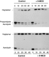
β-MCD does not affect oligomerization, but dramatically accelerates protoxin processing. BHK monolayers were treated with β-MCD (10 mM in IM at 37°C for 1 h), then incubated with either 125I-proaerolysin (a) or trypsin-activated 125I-aerolysin (b; 0.4 nM) for 1 h at 4°C and subsequently incubated at 37°C for various times (indicated in minutes on the figure). PNSs were prepared and analyzed by SDS-PAGE, followed by autoradiography (20 μg of protein were loaded per lane). After β-MCD treatment, conversion of proaerolysin into aerolysin was dramatically accelerated.
Figure 7
Figure 7
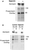
Effects of saponin treatment on aerolysin binding and receptor distribution. a, BHK monolayers were left untreated or treated with saponin (0.4% at 4°C for 1 h) and then incubated with 125I-proaerolysin (0.4 nM) for 1 h at 4°C. PNSs were prepared and analyzed by SDS-PAGE, followed by autoradiography, to detect bound toxin; or by toxin overlay, followed by autoradiography, to detect the proaerolysin receptors (20 μg of protein were loaded per lane). All bands detected by toxin overlay are GPI-anchored proteins and can no longer be detected when cells have been treated with the phosphatidyl inositol-specific phospholipase C (Abrami et al. 1998b). b, BHK monolayers were left untreated or treated with saponin, as in a. Cells were scrapped, pelleted, and resuspended in 1% Triton X-100 at 4°C. Membranes were solubilized by rotary shaking at 4°C for 30 min. The detergent insoluble fractions were obtained by high-speed centrifugation (30 min at 4°C at 55,000 rpm). Detergent insoluble (I) and soluble (S) fractions were analyzed by SDS-PAGE, followed by autoradiography, to detect bound toxin; or by toxin overlay, followed by autoradiography, to detect the proaerolysin receptors. One tenth of each fraction was loaded per lane (corresponding to ∼80 μg of the Triton X-100 soluble fraction) to analyze the recovery of the toxin and its receptors.
Figure 8
Figure 8
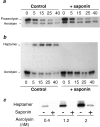
Saponin inhibits the kinetics of aerolysin oligomerization. BHK monolayers were left untreated or treated with saponin (0.4% at 4°C for 1 h). Cells were then incubated with 125I-proaerolysin (0.4 nM; a) or in vitro trypsin-activated 125I-aerolysin (0.4 nM; b) for 1 h at 4°C, washed, and further incubated for various times at 37°C (indicated in minutes on the figure). PNSs were prepared and analyzed by SDS-PAGE, followed by autoradiography (20 μg of protein were loaded per lane). After prolonged incubation at 37°C, a slight loss of cell-bound toxin is observed in saponin-treated cells for reasons that remain to be established. c, BHK monolayers were left untreated or treated with saponin as in a, and then incubated for 1 h at 4°C with different concentrations of in vitro trypsin-activated 125I-aerolysin. At all concentrations tested, binding of aerolysin to the cells was specific. Cells were then washed and further incubated for 25 min at 37°C. PNSs were prepared and analyzed by SDS-PAGE, followed by autoradiography.

References

    1. Abrami L., Fivaz M., Decroly E., Seidah N.G., François J., Thomas G., Leppla S., Buckley J.T., van der Goot F.G. The pore-forming toxin proaerolysin is processed by furin J. Biol. Chem 273 1998. 32656 32661a - PubMed
    1. Abrami L., Fivaz M., Glauser P.-E., Parton R.G., van der Goot F.G. A pore-forming toxin interacts with a GPI-anchored protein and causes vacuolation of the endoplasmic reticulum J. Cell Biol. 140 1998. 525 540b - PMC - PubMed
    1. Adam, G., and M. Delbrück. 1968. Structural Chemistry and Molecular Biology. A. Rich and N. Davidson, editors. W.H. Freeman & Co., NY.
    1. Bhakdi S., Bayley H., Valeva A., Walev I., Walker B., Kehoe M., Palmer M. Staphylococcal alpha-toxin, streptolysin-O, and Escherichia coli hemolysinprototypes of pore-forming bacterial cytolysins. Arch. Microbiol. 1996;165:73–79. - PubMed
    1. Bray D., Levin M.D., Morton-Firth C.J. Receptor clustering as a cellular mechanism to control sensitivity. Nature. 1998;393:85–88. - PubMed

Publication types

MeSH terms