. 1999 Oct 4;190(7):915-22.
doi: 10.1084/jem.190.7.915.

Absence of epithelial immunoglobulin A transport, with increased mucosal leakiness, in polymeric immunoglobulin receptor/secretory component-deficient mice

Affiliations

Absence of epithelial immunoglobulin A transport, with increased mucosal leakiness, in polymeric immunoglobulin receptor/secretory component-deficient mice

F E Johansen et al. J Exp Med. .

Abstract

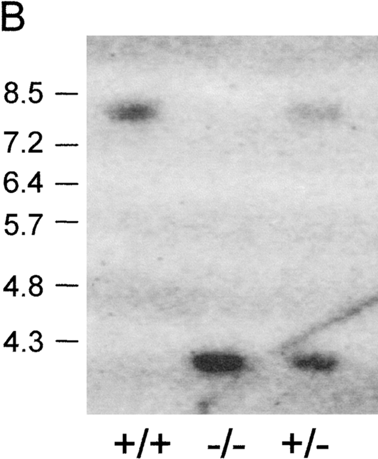
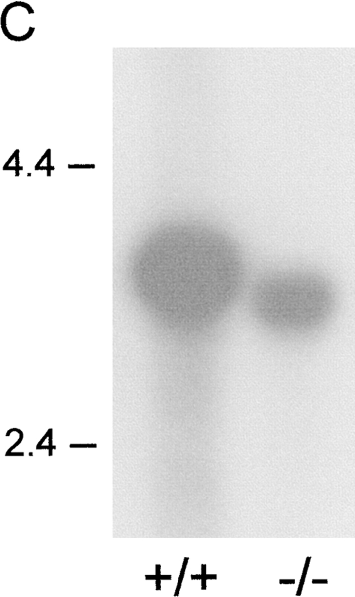
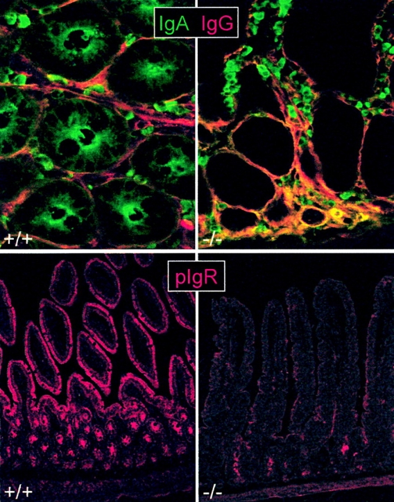
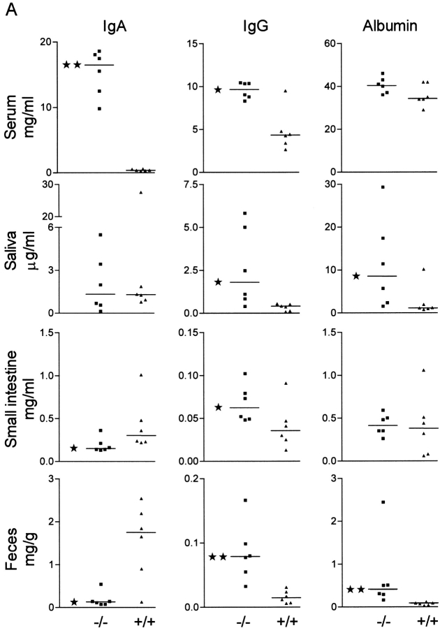
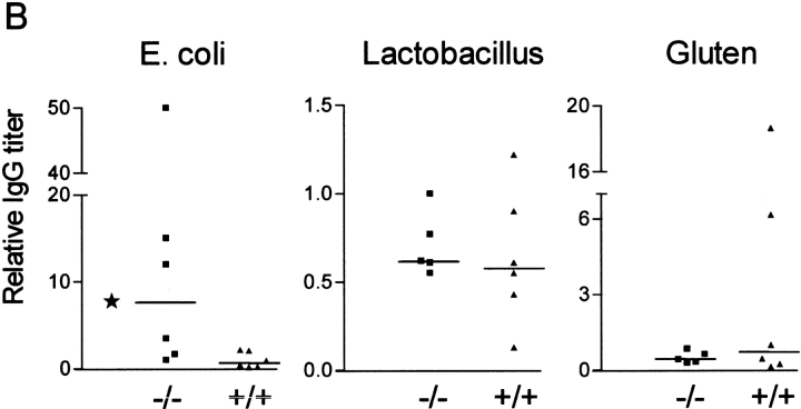
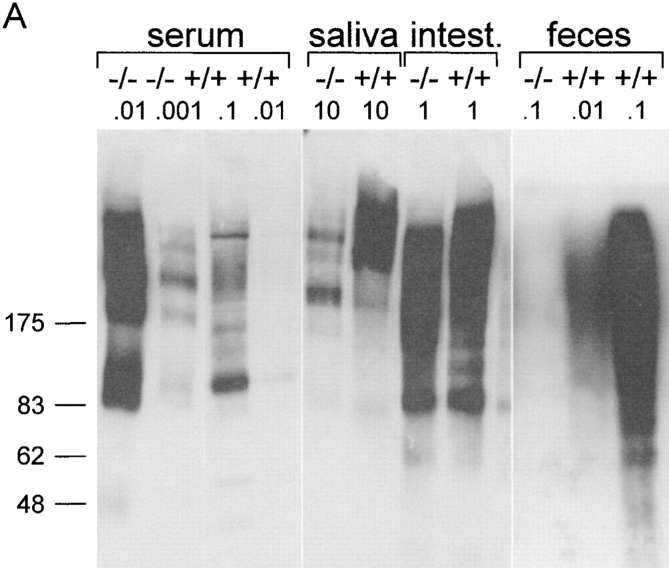
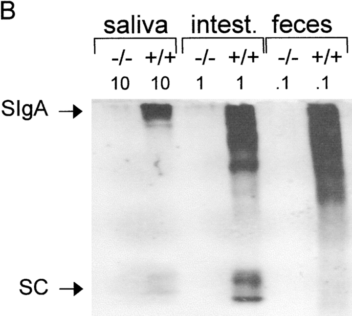
Mucosal surfaces are protected specifically by secretory immunoglobulin A (SIgA) and SIgM generated through external translocation of locally produced dimeric IgA and pentameric IgM. Their active transport is mediated by the epithelial polymeric Ig receptor (pIgR), also called the transmembrane secretory component. Paracellular passive external transfer of systemic and locally produced antibodies also provides mucosal protection, making the biological importance of secretory immunity difficult to assess. Here we report complete lack of active external IgA and IgM translocation in pIgR knockout mice, indicating no redundancy in epithelial transport mechanisms. The knockout mice were of normal size and fertility but had increased serum IgG levels, including antibodies to Escherichia coli, suggesting undue triggering of systemic immunity. Deterioration of their epithelial barrier function in the absence of SIgA (and SIgM) was further attested to by elevated levels of albumin in their saliva and feces, reflecting leakage of serum proteins. Thus, SIgA did not appear to be essential for health under the antigen exposure conditions of these experimental animals. Nevertheless, our results showed that SIgA contributes to maintenance of mucosal homeostasis. Production of SIgA might therefore be a variable in the initiation of human immunopathology such as inflammatory bowel disease or gluten-sensitive enteropathy.


Figures

Figure 1
Figure 1
Generation of pIgR−/− mice. (A) The PIGR locus and gene targeting strategy. A neoR cassette was inserted in exon 3, disrupting the noncovalent pIg-binding site, and a herpes simplex virus thymidine kinase gene was inserted downstream for negative selection of nonhomologous recombinants. (B) Southern blot of tail DNA from wild-type, heterozygote, and pIgR−/− (+/+, +/−, and −/−, respectively) mice probed with the 1.4-kb NcoI fragment indicated in A. (C) Northern blot of RNA extracted from small intestines of +/+ and −/− mice probed with murine pIgR cDNA (gift from C. Kaetzel).
Figure 1
Figure 1
Generation of pIgR−/− mice. (A) The PIGR locus and gene targeting strategy. A neoR cassette was inserted in exon 3, disrupting the noncovalent pIg-binding site, and a herpes simplex virus thymidine kinase gene was inserted downstream for negative selection of nonhomologous recombinants. (B) Southern blot of tail DNA from wild-type, heterozygote, and pIgR−/− (+/+, +/−, and −/−, respectively) mice probed with the 1.4-kb NcoI fragment indicated in A. (C) Northern blot of RNA extracted from small intestines of +/+ and −/− mice probed with murine pIgR cDNA (gift from C. Kaetzel).
Figure 2
Figure 2
Lack of epithelial IgA transport in pIgR−/− mice. Immunofluorescence staining of IgA, IgG, and pIgR/SC in tissue sections from the small intestine of knockout (−/−; right panels) and wild-type (+/+; left panels) mice. Top panels: paired staining of IgA (fluorescein, green) and IgG (Texas Red) in cross section of crypt region. Note lack of IgA in epithelium and increased level of interstitial IgA in pIgR−/− mice (seen as yellow color due to mixing of green with red). Bottom panels: SC staining at low magnification showing abundant pIgR/SC expression in both crypt and villous epithelium of wild-type mice but only occasional faint staining deep in the crypts of pIgR−/− mice. Small intestinal mucosa is shown at full height (note obliquely cut villi in left panel due to tilted tissue orientation).
Figure 3
Figure 3
Altered Ig and albumin levels in serum and secretions of pIgR−/− mice. (A) Levels of IgA, IgG, and albumin in pIgR−/− (−/−, ▪) and wild-type (+/+, ▴) mice were measured by ELISA in serum, saliva, mucus extract from the small intestine, and extract of feces. Statistical analysis was performed with the Mann-Whitney test (★, P < 0.05 and ★★, P < 0.005). (B) Relative titers of serum IgG antibodies against E. coli, Lactobacillus, or gluten.
Figure 3
Figure 3
Altered Ig and albumin levels in serum and secretions of pIgR−/− mice. (A) Levels of IgA, IgG, and albumin in pIgR−/− (−/−, ▪) and wild-type (+/+, ▴) mice were measured by ELISA in serum, saliva, mucus extract from the small intestine, and extract of feces. Statistical analysis was performed with the Mann-Whitney test (★, P < 0.05 and ★★, P < 0.005). (B) Relative titers of serum IgG antibodies against E. coli, Lactobacillus, or gluten.
Figure 4
Figure 4
Increased pIgA serum level but lack of SIgA in pIgR−/− mice. Western blot of IgA in serum and secretions from pIgR−/− (−/−) and wild-type (+/+) mice. The indicated amounts of samples (in microliters for secretions and micrograms dry weight for feces) were separated by SDS-PAGE, transferred to nitrocellulose, and probed with antibody to α chain (A) or to SC as a marker of SIgA (B). The positions of migration of purified human SC and SIgA are indicated. Intest., intestine.
Figure 4
Figure 4
Increased pIgA serum level but lack of SIgA in pIgR−/− mice. Western blot of IgA in serum and secretions from pIgR−/− (−/−) and wild-type (+/+) mice. The indicated amounts of samples (in microliters for secretions and micrograms dry weight for feces) were separated by SDS-PAGE, transferred to nitrocellulose, and probed with antibody to α chain (A) or to SC as a marker of SIgA (B). The positions of migration of purified human SC and SIgA are indicated. Intest., intestine.

Similar articles

Cited by

References

    1. Brandtzaeg P. History of oral tolerance and mucosal immunity. Ann. NY Acad. Sci. 1996;778:1–27. - PubMed
    1. Mestecky J., Moro I., Underdown B. Mucosal immunoglobulins. In: Mestecky J., Bienstock J., McGhee J., Lamm M., Strober W., Ogra P., editors. Mucosal Immunology. Academic Press; San Diego, CA: 1999. p. 133.
    1. Stokes C.R., Soothill J.F., Turner M.W. Immune exclusion is a function of IgA. Nature. 1975;255:745–746. - PubMed
    1. Mazanec M.B., Kaetzel C.S., Lamm M.E., Fletcher D., Nedrud J.G. Intracellular neutralization of virus by immunoglobulin A antibodies. Proc. Natl. Acad. Sci. USA. 1992;89:6901–6905. - PMC - PubMed
    1. Mazanec M.B., Nedrud J.G., Kaetzel C.S., Lamm M.E. A three-tiered view of the role of IgA in mucosal defense. Immunol. Today. 1993;14:430–435. - PubMed

Publication types

MeSH terms