. 1999 Oct 4;190(7):923-34.
doi: 10.1084/jem.190.7.923.

Cytokine-activated endothelial cells delay neutrophil apoptosis in vitro and in vivo. A role for granulocyte/macrophage colony-stimulating factor

Affiliations

Cytokine-activated endothelial cells delay neutrophil apoptosis in vitro and in vivo. A role for granulocyte/macrophage colony-stimulating factor

A Coxon et al. J Exp Med. .

Abstract

The activation of endothelium is important in recruiting neutrophils to sites of inflammation and in modulating their function. We demonstrate that conditioned medium from cultured, activated endothelial cells acts to significantly delay the constitutive apoptosis of neutrophils, resulting in their enhanced survival and increased phagocytic function. The antiapoptotic activity is, in part, attributable to granulocyte/macrophage colony-stimulating factor (GM-CSF) secreted by activated endothelial cells. The in vivo relevance of these findings was investigated in a cytokine-induced model of acute meningitis in mice. Peripheral blood neutrophils (PBNs) from mice with meningitis exhibited a delay in apoptosis compared with untreated mice. Furthermore, neutrophils recovered from the inflamed cerebrospinal fluid (CSF) exhibited enhanced survival compared with neutrophils isolated from the peripheral blood of the same animals. In unchallenged GM-CSF-deficient mice, the apoptosis of circulating PBNs was similar to wild-type animals; however, after cytokine-induced meningitis, the delay in neutrophil apoptosis typically observed in wild-type mice was attenuated. In contrast, the apoptosis of neutrophils recovered from the CSF of mice of both genotypes was comparable. Taken together, these studies suggest that neutrophil apoptosis is regulated during an inflammatory response, in both intravascular and extravascular compartments. GM-CSF released by activated endothelium can act to increase neutrophil survival and function in the peripheral blood, whereas other factor(s) appear to perform this function in the extravascular space.


Figures

Figure 1
Figure 1
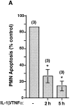
PBNs and neutrophils extravasated into the CSF of mice after cytokine-induced meningitis have delayed apoptosis, and an activity released into the CSF is responsible for this delay. 129Sv wild-type mice were subjected to cytokine-induced meningitis by injection of IL-1β/TNF-α by lumbar puncture or were left untreated (A). After 4 h, CSF containing leukocyte infiltrate was collected from each mouse (n = 6). PMNs represented >90% of the cells present. Peripheral blood was pooled from two groups of two mice which had been subjected to meningitis, and two groups of two untreated mice. PMNs from each CSF (PMN/CSF) and peripheral blood neutrophil (PBNs) samples were cultured in duplicate, and neutrophil apoptosis was assessed at the indicated times (*P < 0.005, # P < 0.02 at both time points). (B) Representative cytospins of the 5-h time point of PMNs from the CSF, and PBNs from cytokine-injected mice are shown. Apoptotic neutrophils are indicated by arrows. In a separate experiment, CSF was harvested from untreated mice and from mice 2 h after induction of meningitis (C). The isolated CSF was centrifuged to remove any cells that might be present; importantly, at this early time point very few neutrophils are present in the CSF. PBNs from untreated mice were cultured in the CSF or medium for 4 h. The neutrophils were then diluted with medium, and apoptosis was assessed after an additional 5 h. n = 3 individual experiments; *P < 0.05 compared with CSF/untreated mice.
Figure 2
Figure 2
Cytokine-activated endothelial cells significantly delay neutrophil apoptosis, and inhibition of de novo synthesis abrogates expression of the antiapoptotic activity. (A) Neutrophils were allowed to transmigrate through untreated (−) or IL-1β/TNF-α–activated (Cyt.EC) HUVEC monolayers. Buffer alone or buffer plus the chemoattractants FMLP or LTB4 were included in the bottom chamber of the transwell. Neutrophils incubated with untreated HUVECs transmigrated in response to FMLP in the bottom chamber. Control neutrophils were incubated in the presence or absence of the respective chemoattractant, without endothelial cells. (B) Neutrophils were cocultured with HUVEC monolayers for 1 h after HUVECs were stimulated with IL-1β/TNF-α for the indicated times. 2+4 wo indicates that 2 h cytokine–treated HUVECs were washed, incubated for 4 h without cytokines, and then incubated with neutrophils for 1 h. For A and B, *P < 0.005 and # P < 0.05 compared with untreated HUVEC monolayers. (C) Endothelial cells were pretreated with 12 μM actinomycin D (+ Act.D) or 50 μg/ml cycloheximide (+ CX) for 1 h, and IL-1β/TNF-α was added for an additional 1 h. The monolayer was washed, and neutrophils were added for 1 h. # P < 0.05 compared with IL-1β/TNF-α. For A, B, and C, neutrophils were recovered after transmigration or coculture, and apoptosis was assessed 6 h after culturing in fresh medium. Data are expressed as percentage of apoptosis of control neutrophils (% control) to account for the donor variability in the amount of spontaneous apoptosis after 6 h. n = 4 experiments.
Figure 2
Figure 2
Cytokine-activated endothelial cells significantly delay neutrophil apoptosis, and inhibition of de novo synthesis abrogates expression of the antiapoptotic activity. (A) Neutrophils were allowed to transmigrate through untreated (−) or IL-1β/TNF-α–activated (Cyt.EC) HUVEC monolayers. Buffer alone or buffer plus the chemoattractants FMLP or LTB4 were included in the bottom chamber of the transwell. Neutrophils incubated with untreated HUVECs transmigrated in response to FMLP in the bottom chamber. Control neutrophils were incubated in the presence or absence of the respective chemoattractant, without endothelial cells. (B) Neutrophils were cocultured with HUVEC monolayers for 1 h after HUVECs were stimulated with IL-1β/TNF-α for the indicated times. 2+4 wo indicates that 2 h cytokine–treated HUVECs were washed, incubated for 4 h without cytokines, and then incubated with neutrophils for 1 h. For A and B, *P < 0.005 and # P < 0.05 compared with untreated HUVEC monolayers. (C) Endothelial cells were pretreated with 12 μM actinomycin D (+ Act.D) or 50 μg/ml cycloheximide (+ CX) for 1 h, and IL-1β/TNF-α was added for an additional 1 h. The monolayer was washed, and neutrophils were added for 1 h. # P < 0.05 compared with IL-1β/TNF-α. For A, B, and C, neutrophils were recovered after transmigration or coculture, and apoptosis was assessed 6 h after culturing in fresh medium. Data are expressed as percentage of apoptosis of control neutrophils (% control) to account for the donor variability in the amount of spontaneous apoptosis after 6 h. n = 4 experiments.
Figure 3
Figure 3
Conditioned medium from cytokine-stimulated HUVECs significantly delays neutrophil apoptosis. (A) HUVECs were left untreated (−) or were treated with IL-1β/TNF-α for the indicated times, washed, and incubated with buffer alone for 1 h. The conditioned buffer was removed and incubated with neutrophils for 1 h. The neutrophils were then put into fresh medium, and apoptosis was assessed after 6 h. (B) Conditioned medium was collected from untreated (unt. EC) and 1.5-h cytokine-activated (IL-1/TNF EC) HUVECs, as described above. In contrast to conditions in A, the neutrophils were cultured for 18–24 h in the continuous presence of conditioned medium or medium alone, and percentage of apoptosis was assessed. The number of experiments performed is shown in parentheses. (C) Representative cytospins are shown, and apoptotic cells are indicated by arrows. (D) A representative FACS® analysis of samples in B that were stained with FITC–Annexin V, which preferentially binds to apoptotic cells. *P < 0.005 compared with IL-1β/TNF-α (A) or medium alone (B).
Figure 3
Figure 3
Conditioned medium from cytokine-stimulated HUVECs significantly delays neutrophil apoptosis. (A) HUVECs were left untreated (−) or were treated with IL-1β/TNF-α for the indicated times, washed, and incubated with buffer alone for 1 h. The conditioned buffer was removed and incubated with neutrophils for 1 h. The neutrophils were then put into fresh medium, and apoptosis was assessed after 6 h. (B) Conditioned medium was collected from untreated (unt. EC) and 1.5-h cytokine-activated (IL-1/TNF EC) HUVECs, as described above. In contrast to conditions in A, the neutrophils were cultured for 18–24 h in the continuous presence of conditioned medium or medium alone, and percentage of apoptosis was assessed. The number of experiments performed is shown in parentheses. (C) Representative cytospins are shown, and apoptotic cells are indicated by arrows. (D) A representative FACS® analysis of samples in B that were stained with FITC–Annexin V, which preferentially binds to apoptotic cells. *P < 0.005 compared with IL-1β/TNF-α (A) or medium alone (B).
Figure 4
Figure 4
Effect of immunodepleting conditioned medium of GM-CSF on PMN apoptosis. Conditioned medium (CM) from IL-1β/TNF-α–treated HUVECs was immunodepleted of GM-CSF using neutralizing polyclonal antibodies (CM αGM-CSF). A rabbit IgG was used as negative control (CM IgG). PMNs were incubated for 18 h in the conditioned medium, and the percentage of apoptotic PMNs was assessed. # P < 0.05 compared with medium alone, and *P < 0.05 compared with CM and CM/IgG. The amount of GM-CSF present in the conditioned medium before and after immunodepletion was assessed by ELISA, and the average (pg/ml) is shown below the respective conditions. n = 3 experiments.
Figure 5
Figure 5
Mice deficient in GM-CSF have less of a delay in PBN apoptosis after cytokine-induced meningitis than wild-type mice. Neutrophils were isolated from the CSF and peripheral blood of eight wild-type (WT) and eight GM-CSF–deficient (GM-CSF−/−) mice, 4 h after cytokine-induced meningitis. The harvested neutrophils were then placed in culture, and apoptosis was assessed on cytospins at the indicated time points. *P < 0.05, # P < 0.002.

References

    1. Bicknell S., van Eeden S., Hayashi S., Hards J., English D., Hogg J.C. A non-radioisotopic method for tracing neutrophils in vivo using 5′-bromo-2′-deoxyuridine. Am. J. Respir. Cell Mol. Biol. 1994;10:16–23. - PubMed
    1. Savill J., Haslett C. Fate of neutrophils. In: Hellewell P.G., Williams T.J., editors. Immunopharmacology of Neutrophils. Academic Press, Inc; San Diego: 1994. pp. 295–314.
    1. Whyte M.K.B., Meagher L.C., MacDermot J., Haslett C. Impairment of function in aging neutrophils is associated with apoptosis. J. Immunol. 1993;150:5124–5134. - PubMed
    1. Dransfield I., Stocks S.C., Haslett C. Regulation of cell adhesion expression and function associated with neutrophil apoptosis. Blood. 1995;85:3264–3273. - PubMed
    1. Colotta F., Re F., Polentarutti N., Sozzani S., Mantovani A. Modulation of granulocyte survival and programmed cell death by cytokines and bacterial products. Blood. 1992;80:2012–2020. - PubMed

Publication types

MeSH terms