. 1999 Oct;104(7):855-64.
doi: 10.1172/JCI7691.

Induction of the hair growth phase in postnatal mice by localized transient expression of Sonic hedgehog

Affiliations

Induction of the hair growth phase in postnatal mice by localized transient expression of Sonic hedgehog

N Sato et al. J Clin Invest. 1999 Oct.

Abstract

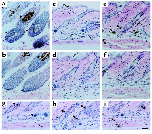
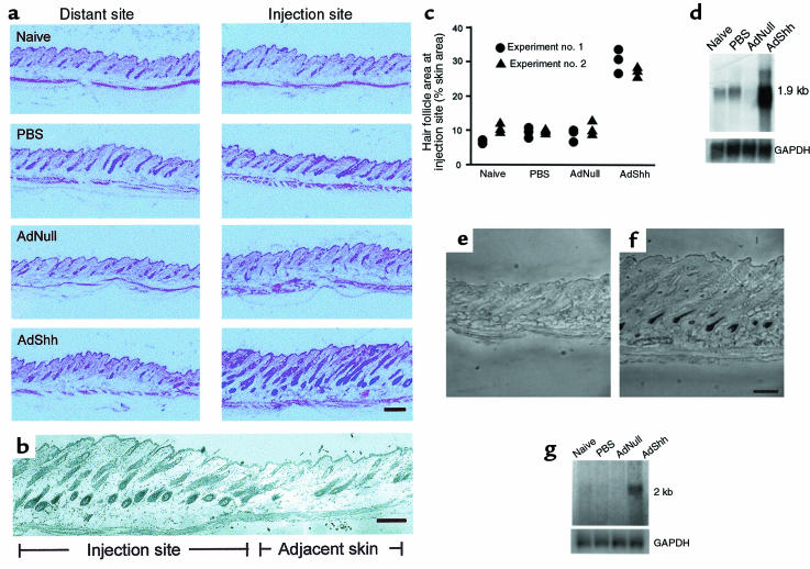
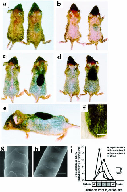
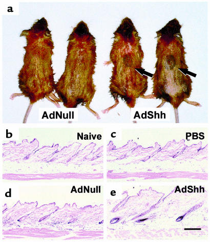
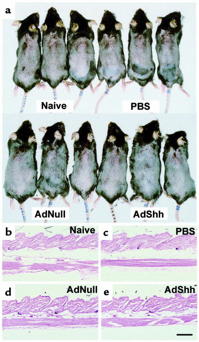
Hair follicles form in prenatal skin and mature in the postnatal period, establishing a growth cycle in 3 phases: telogen (resting), anagen (growth), and catagen (regression). Based on the knowledge that Sonic hedgehog (Shh) expression is necessary for the embryonic development of hair follicles, and that anagen in the postnatal cycling follicle has morphologic similarities to the epithelial invagination process in embryonic skin, we hypothesized that localized, but transient, enhanced expression of the Shh gene in postnatal skin would accelerate initiation of anagen in the hair follicle cycle, with concomitant accelerated hair growth. To assess this concept, an E1(-) adenovirus vector, AdShh, was used to transfer the murine Shh cDNA to skin of postnatal day 19 C57BL/6 mice. The treated skin showed increased mRNA expression of Shh, Patched (the Shh receptor), and Gli1 (a transcription factor in the Shh pathway). In mice receiving AdShh, but not in controls, acceleration into anagen was evident, since hair follicle size and melanogenesis increased and the hair-specific keratin ghHb-1 and the melanin synthesis-related tyrosinase mRNAs accumulated. Finally, C57BL/6 mice showed marked acceleration of the onset of new hair growth in the region of AdShh administration to skin 2 weeks after treatment, but not in control vector-treated or untreated areas. After 6 months, AdShh-treated skin showed normal hair and normal skin morphology. Together, these observations are consistent with the concept that upregulation of Shh activity in postnatal skin functions as a biologic switch that induces resting hair follicles to enter anagen with consequent hair growth.


Figures

Figure 1
Figure 1
Functional expression of Shh by administration of AdShh in vitro and in vivo in postnatal day 19 C57BL/6 mice. (a) Shh mRNA after in vitro infection of A549 epithelial cells with AdShh. A549 cells (106) were infected (20 moi) with the AdNull control vector or AdShh. After 2 days, RNA was analyzed by Northern blot with a 32P-labeled Shh cDNA probe. Uninfected cells (naive) and AdNull-infected cells were negative, whereas AdShh-infected cells contained a transcript of approximately 3.0 kb corresponding to the vector-encoded Shh transgene. Equal RNA loading was confirmed by analysis of GAPDH mRNA. (b) Shh protein expressed in vitro. Protein from A549 cells infected with AdNull or AdShh as described in a was analyzed by immunoblot for Shh protein. AdShh-infected, but not naive or AdNull-infected, A549 cells showed a 19-kDa immunoreactive band with anti-Shh. Equal protein loading was confirmed by Coomassie blue staining of an identically loaded gel (not shown). (c) Time course demonstrating expression of Shh, Ptc, and Gli1 mRNA in vivo. Dorsal skin was collected from naive C57BL/6 mice or C57BL/6 mice at postnatal days 19, 22, 24, 26, and 33 after intradermal injection on day 19 with AdNull or AdShh (108 PFU for either vector). Skin was analyzed for Shh, Ptc, or Gli1 mRNA by Northern analysis on experimental days 0, 3, 5, 7, and 14. Expression of the 3.0-kb mRNA vector-encoded transcript (upper arrow) and the 2.6-kb endogenous Shh transcript (lower arrows) was detected in AdShh-injected mice, but the vector-derived transcript temporally preceded the endogenous transcript. Marked upregulation of Ptc and Gli1 gene expression was detected in AdShh-injected mice on experimental days 3 and 5, when low levels of expression of these genes were seen in naive and AdNull-treated mice. Equal RNA loading was confirmed by analysis of GAPDH mRNA.
Figure 2
Figure 2
Localization of Shh expression in skin by in situ hybridization before and after administration of AdShh. Paraffin sections of mouse skin during its natural anagen period (postnatal day 33) compared with postnatal day 22 naive mouse skin or skin of mice injected on postnatal day 19 with PBS, AdNull, or AdShh. The sections were analyzed by in situ hybridization using [33P]UTP-labeled antisense and sense Shh riboprobes. After hybridization, sites of probe binding were identified using a photographic emulsion, and tissue was stained with hematoxylin and eosin. Arrows indicate positive staining for Shh mRNA. Closed arrowheads indicate melanosomes in the hair follicle. Open arrowheads indicate melanin in hair shafts. (a) Anagen skin from a naive 33-day-old mouse (positive control) hybridized with an antisense Shh complementary to Shh mRNA. (b) Adjacent tissue section of anagen skin hybridized with a sense Shh probe. (c) Naive postnatal day 22 skin hybridized with an antisense Shh probe. (d) Postnatal day 22 skin 3 days after injection with 108 PFU of AdNull hybridized with an antisense Shh probe. (e) Postnatal day 22 skin 3 days after injection of 108 PFU of AdShh hybridized with an antisense Shh probe. (f) Postnatal day 22 skin 3 days after injection with 108 PFU of AdShh hybridized with a sense Shh probe. (gi) High-magnification examples of cells in AdShh-injected mouse skin hybridized with antisense Shh probe. Scale bar: 50 μm.
Figure 3
Figure 3
Induction of hair follicle growth and melanogenesis after intradermal administration of AdShh. AdShh, AdNull, or PBS was administered to the dorsal skin of postnatal day 19 C57BL/6 mice as in Figure 1c, and analyses were performed on postnatal day 26. (a) Histologic evaluation 7 days after vector administration. The AdShh-injected skin has increased depth of the dermal layer, increased follicle length, and increased follicle area compared with the naive, PBS, and AdNull controls. The changes associated with AdShh injection were limited to the area of injection (injection site vs. distant site [control]). (b) Histologic evaluation of the border of the injection site 7 days after vector administration. Hair follicles in the injection site are in late anagen; hair follicles in adjacent skin are in early anagen. A corresponding change in skin thickness was noted. (c) Quantitation of follicle area as a percentage of the total dermal/epidermal area using digital imaging and pixel area integration. Each data point represents an area measurement from a representative field. Data from 3 fields are shown per mouse; data from 2 mice are shown per treatment. (d) Northern analysis showing enhanced expression of the hair-specific keratin ghHb-1 gene induced by AdShh in vivo relative to the naive, PBS, and AdNull controls. Equal RNA loading shown by probing for GAPDH mRNA. (e and f) Melanin expression in hair follicle. Accumulation of melanin was evaluated by bright-field microscopy of unstained paraffin sections. Skin at the site of injection of AdShh (f), but not AdNull-injected skin (e), showed increased melanin in hair follicle bulbs. Skin of PBS-injected and naive control mice was similar to skin of AdNull-injected mice (not shown). (g) Northern analysis showing upregulation of melanogenesis-related tyrosinase gene expression after administration of AdShh relative to the naive, PBS, and AdNull controls. Equal RNA loading was confirmed by probing for GAPDH mRNA. Scale bars: 200 μm.
Figure 4
Figure 4
Hair growth in C57BL/6 mice after intradermal administration of AdShh during first telogen. AdShh, AdNull, or PBS was administered to the dorsal skin of postnatal day 19 C57BL/6 mice. Five days after administration, dorsal hair was bleached with blonde hair dye to provide contrast for assessing new growth of the natural black hair of the mice. On day 7, the dorsal hair was clipped. (a and b) Mice at day 7 after administration of AdShh. Shown are 2 example pairs. The injection site in AdNull-treated animals was indistinct (left-side mice), whereas melanogenesis was evident at the site of AdShh injection in AdShh-treated mice (right-side mice). (c and d) Mice at day 14 after administration of AdShh. Shown are 2 example pairs. The injection site in AdNull-treated animals was indistinct (left-side mice). New hair growth is seen (note black color relative to preexisting dyed hair) at the site of AdShh injection (right-side mice). (e) Lateral aspect of day 14 AdShh-treated mouse showing length of hair growth at injection site. (f) High magnification of injection site showing new black hair and preexisting blonde-dyed hair. Scale bar: 2 mm. (g) Scanning electron microscopic analysis of normal C57BL/6 mouse hair shaft. (h) Scanning electron microscopic analysis of C57BL/6 mouse hair shaft induced by intradermal injection of AdShh (day 14 after vector administration). Scale bar: 10 μm. (i) Spatial distribution of transgene expression. Adβgal (108 PFU), an E1E3 Ad vector expressing the Escherichia coli βgal marker gene, was injected intradermally on the dorsal surface of postnatal day 19 C57BL/6 mice. On day 0 and on day 2, a single strip of dorsal skin along the cephalocaudal axis was harvested, divided into 5 equal 2.5-mm segments (see diagram below graph; 0 = site of infection), and assayed for βgal activity (data from 3 animals are shown). The gray area corresponds to the size and position of the wheal (7–10 mm in diameter) formed at the site of injection. Data are expressed as βgal activity per mg protein. The anatomic distribution of the marker gene is similar to that of the melanogenesis and new hair growth observed in b and c.
Figure 5
Figure 5
Hair growth in C57BL/6 mice after intradermal administration of AdShh during second telogen. AdShh, AdNull, or PBS was administered to the dorsal skin of 8-week-old C57BL/6 mice. Mice were dyed and clipped as described in Figure 3, and were evaluated on experimental day 10 after vector administration for melanogenesis. Dorsal skin was subsequently collected, embedded in paraffin, and sectioned along the cephalocaudal axis. (a) Melanogenesis induced by AdShh. AdShh-treated mice (right pair of mice) but not AdNull-treated mice (left pair of mice) showed melanogenesis at the site of injection 7 days after treatment. (be) Histologic evaluation of area of AdShh injection. (b) Naive mouse skin. (c) PBS-injected mouse skin. (d) AdNull-injected mouse skin. (e) AdShh-injected mouse skin. Note the increased thickness and increased area of hair follicles similar to that in Figure 3. Scale bar: 200 μm.
Figure 6
Figure 6
Macroscopic and histologic assessment of AdShh-injected mice 6 months after administration of AdShh. (a) Macroscopic assessment of PBS-, AdNull-, and AdShh-injected mice. For long-term studies, mice were not exposed to hair dye, but were clipped once at the time of vector administration. No differences were noted among groups. (be) Histologic assessment. Shown are representative sections from skin of a naive mouse and of PBS-, AdNull-, and AdShh-injected mice 6 months after administration. No abnormalities were observed at the injection site at this time. Three mice were examined for each treatment. Scale bar: 200 μm.

Comment in

Similar articles

Cited by

References

    1. Hardy MH. The secret life of the hair follicle. Trends Genet. 1992;8:55–61. - PubMed
    1. Oro AE, Scott MP. Splitting hairs: dissecting roles of signaling systems in epidermal development. Cell. 1998;95:575–578. - PubMed
    1. Sengel, P. 1976. Morphogenesis of skin. Cambridge University Press. Cambridge, United Kingdom. pp. 20–29.
    1. Wessells NK, Rommens JM. Nonproliferation in dermal condensations of mouse vibrissae and pelage hairs. Dev Biol. 1965;12:419–433. - PubMed
    1. Bertolino, A.P., Klein, L.M., and Freedberg, I.M. 1993. Biology of hair follicles. In Dermatology in general medicine. T.B. Fitzpatrick, A.Z. Eisen, K. Wolff, I.M. Freedberg, and K.F. Austen, editors. McGraw-Hill Inc. New York, NY. 289–293.

Publication types