. 1999 Oct;104(7):875-84.
doi: 10.1172/JCI7270.

Epithelial P2X purinergic receptor channel expression and function

Affiliations

Epithelial P2X purinergic receptor channel expression and function

A L Taylor et al. J Clin Invest. 1999 Oct.

Abstract

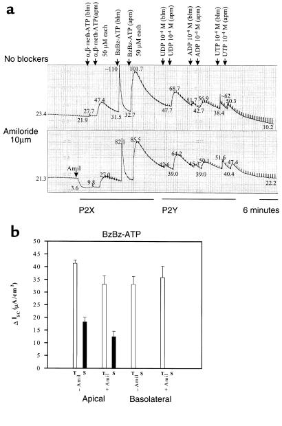
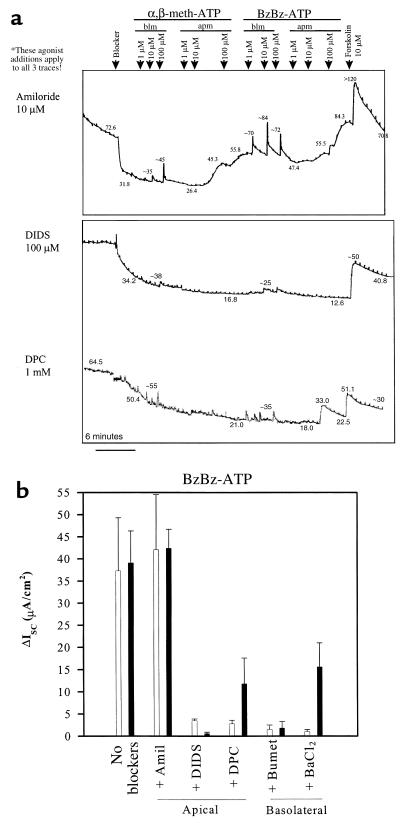
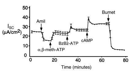
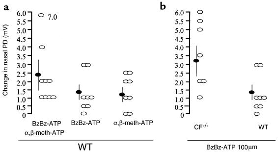
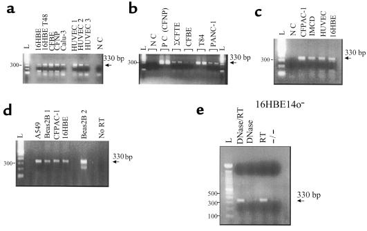
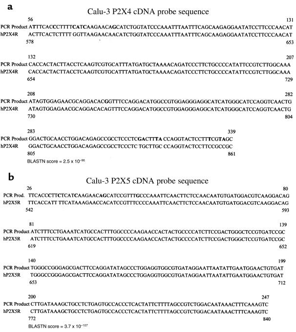
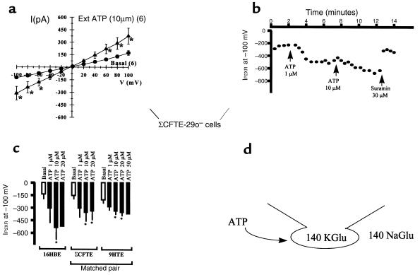
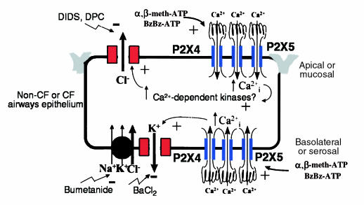
P2X purinergic receptor (P2XR) channels bind ATP and mediate Ca(2+) influx--2 signals that stimulate secretory Cl(-) transport across epithelia. We tested the hypotheses that P2XR channels are expressed by epithelia and that P2XRs transduce extracellular ATP signals into stimulation of Cl(-) transport across epithelia. Electrophysiological data and mRNA analysis of human and mouse pulmonary epithelia and other epithelial cells indicate that multiple P2XRs are broadly expressed in these tissues and that they are active on both apical and basolateral surfaces. Because P2X-selective agonists bind multiple P2XR subtypes, and because P2X agonists stimulate Cl(-) transport across nasal mucosa of cystic fibrosis (CF) patients as well as across non-CF nasal mucosa, P2XRs may provide novel targets for extracellular nucleotide therapy of CF.


Figures

Figure 1
Figure 1
Apical (apm) and basolateral (blm) P2X agonists stimulate ISC across mouse tracheal epithelial monolayers grown in primary culture. (a) Parallel recordings with and without apical amiloride are shown. Measured current values (in μA/cm2) are shown at points along each record. P2YR-selective agonists were also tested in these experiments. No washing of agonists was performed. (b) Data summary of transient (T) and prolonged (S) ISC stimulated by the agonist, BzBz-ATP. n = 4 with and without amiloride. All stimulated current was significant (P < 0.005). Similar effects were obtained in experiments with α,β-meth-ATP.
Figure 2
Figure 2
Apical (apm) and basolateral (blm) P2X agonists stimulate ISCCl across mouse tracheal epithelial monolayers grown in primary culture. (a) Parallel recordings in the presence of apical amiloride, apical DIDS, and apical DPC. Forskolin was added as a positive control to stimulate CFTR-dependent ISC and to confirm the integrity of each monolayer. (b) Data summary showing effects of apical and basolateral blockers of ion transport on BzBz-ATP–stimulated ISC. n = 4 with and without amiloride; n = 3 for other blockers. All stimulated current was significant (P < 0.005). Similar effects were obtained in experiments with α,β-meth-ATP. Amil, amiloride; Bumet, bumetanide.
Figure 3
Figure 3
Apical P2X agonists stimulate ISCCl in an additive manner across human non-CF airway epithelial monolayers grown in primary culture. Mean ± SEM of 3 recordings performed on human non-CF airway epithelial monolayers grown in primary culture. A cocktail containing forskolin, IBMX, and cAMP analogue (cAMP) was added as a positive control to stimulate CFTR-dependent ISC and to confirm the integrity of each monolayer. Bumetanide blocked approximately 90% of the stimulated secretory Cl current. The residual current may be amiloride-insensitive Na+ absorptive current (perhaps P2XR current itself). All effects of inhibitors and agonists were significant (P < 0.05).
Figure 4
Figure 4
Mucosal P2X agonists stimulate secretory Cl transport across wild-type and CFTR-knockout mouse nasal epithelia in vivo. (a) Data summary showing individual experiments (open ovals) and the mean ± SEM values (filled ovals) from studies with each P2XR agonist and with both agonists added together as a cocktail. (b) Data summary showing comparison of effects of BzBz-ATP in CFTR-knockout vs. wild-type (WT) mice. Shown are individual trials (open ovals) and mean ± SEM (filled ovals). Steady-state nasal PD values are shown after addition of P2XR agonist in low-Cl solution with amiloride. Change in PD is a depolarization indicative of an increase in Cl permeability; the change was significant in all cases (P < 0.05).
Figure 5
Figure 5
P2XR mRNA is expressed in human CF and non-CF airway epithelial cell lines and in primary cultures. (a) P2XR PCR products from 16HBE14o non-CF human bronchial epithelial cells, CFBE41o CF human bronchial epithelial cells, ΣCFTE-29o CF human tracheal epithelial cells (shown in b), a CFNP epithelial primary culture, Calu-3 non-CF human submucosal gland serous cells, and human umbilical vein endothelial cells (HUVEC). NC, no cDNA (control). (b) Comparison of positive airway epithelial cell reactions to T84 non-CF human colon carcinoma cells and PANC-1 non-CF human pancreatic epithelial cells. (c) Positive reactions from the 16HBE14o sample compared with CFPAC-1 CF human pancreatic epithelial cells, IMCD renal inner medullary collecting-duct cells, and HUVEC. (d) Positive reactions for A549 non-CF human alveolar lung cells and Beas2B non-CF human bronchial epithelial cells vs. control (No RT). Positive reactions for CFPAC-1 and 16HBE14o cells are also shown as positive controls for comparison. Beas2B 1 and Beas2B 2 are two different cDNA samples from the same RNA sample. (e) PCR controls on 16HBE14o total RNA sample. No P2XR PCR product was derived from amplification of total RNA (–/–) or RNase-free, DNase-treated total RNA (DNase), whereas positive reactions occurred in total RNA reverse-transcribed to cDNA with (DNase/RT) and without (RT) DNase treatment. All amplifications of epithelial cell samples were performed 2–3 times. Note: Analysis of the sites that the degenerate P2X PCR primers recognized within the genomic sequence of the mouse P2X3 gene (28) indicated that our primers spanned at least 2 introns; amplification of genomic DNA would produce a PCR product at least 2 kb larger than the 330-bp expected size. Moreover, in the P2X3 genomic sequence, our reverse primer spanned an exon/intron boundary (intron ∼25 kb in size), rendering amplification of genomic DNA improbable.
Figure 6
Figure 6
Representative P2X4 and P2X5 cDNA probe sequences. Nucleotides in bold are differences between our PCR products and the published GenBank sequence. BLASTN score of identity is shown for each sequence. Tables 1 and 2 reflect the number of sequences in all cell models screened that were identified as P2X4 (a) or P2X5 (b). Sequences derived from degenerate RT-PCR of a Calu-3 total RNA sample are shown. A BLASTNscore that is more negative in its exponent reflects a high degree of sequence identity.
Figure 7
Figure 7
Extracellular ATP (Ext ATP) stimulates nonselective cation currents in whole-cell recordings of non-CF and CF airway epithelial cells. (a) I-V plots of basal and ATP-stimulated nonselective cation currents in ΣCFTE-29o cells. n = 6, paired recordings. Values are mean ± SEM (*P < 0.05). (b) Typical time course of sustained P2XR current stimulation with 2 doses of ATP ligand, and reversal with suramin, a P2XR antagonist. (c) Data summary of ATP ligand stimulation of P2XR current in 3 human airway epithelial cell models. n = 4–6 among the 3 airway epithelial cell models. Similar data was obtained using T84 cells (data not shown). (d) Patch-clamp icon illustrates the experimental design, where the pipette (intracellular) solution contains 140 mM potassium gluconate (KGlu) and the bath (extracellular) solution contains 145 mM sodium gluconate (NaGlu). In this design, whole-cell currents are nonselective for cations if the reversal potential is at or near 0 mV. Substitution of Cl with Glu eliminates the chance that ATP agonists would stimulate an epithelial Cl channel, a possibility that is probably based on the preliminary data above. Moreover, Glu also chelates divalent cations, such as Ca2+ and Mg2+, that may block P2XR channels.
Figure 8
Figure 8
Model of P2XR channel expression and function in non-CF or CF airway epithelium. The model is reflective of either a non-CF or CF airways epithelium. P2XR channels are expressed on both the apical and basolateral membranes. They probably form heteromultimeric complexes of P2X4 and P2X5, based on our molecular expression data; however, additional isoforms have been detected in some epithelial cell lines. The electrophysiological and signaling mechanisms whereby P2XRs may stimulate Cl secretion are shown in the apical and basolateral membrane domains. Ca2+-mediated signaling is probable; however, additional unappreciated signaling mechanisms may be involved that are triggered by P2XRs.

References

    1. Abbracchio MP, Burnstock G. Purinoceptors: are there families of P2X and P2Y purinoceptors? Pharmacol Ther. 1994;64:445–475. - PubMed
    1. Barnard EA, Burnstock G, Webb TE. G protein-coupled receptors for ATP and other nucleotides: a new receptor family. Trends Pharmacol Sci. 1994;15:67–70. - PubMed
    1. Buell G, Collo G, Rassendren F. P2X receptors: an emerging channel family. Eur J Neurosci. 1996;8:2221–2228. - PubMed
    1. Bean BP. Pharmacology and electrophysiology of ATP-activated ion channels. Trends Pharmacol Sci. 1992;13:87–90. - PubMed
    1. Parr CE, et al. Cloning and expression of a human P2U nucleotide receptor, a target for cystic fibrosis pharmacotherapy. Proc Natl Acad Sci USA. 1994;91:3275–3279. - PMC - PubMed

Publication types

MeSH terms