

Cavernous sinus and inferior petrosal sinus flow signal on three-dimensional time-of-flight MR angiography
- PMID: 10512234
- PMCID: PMC7657743
Cavernous sinus and inferior petrosal sinus flow signal on three-dimensional time-of-flight MR angiography
Abstract
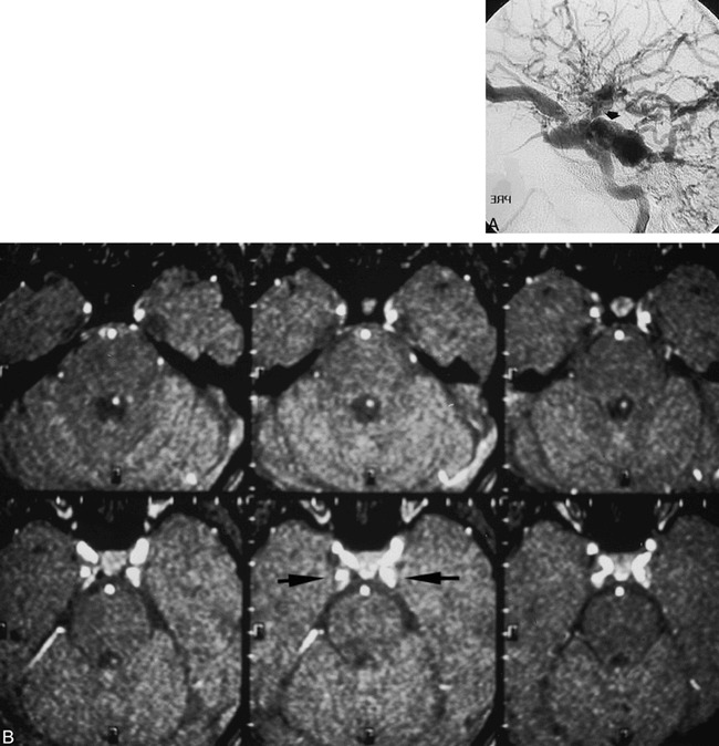
Background and purpose: Venous flow signal in the cavernous sinus and inferior petrosal sinus has been shown on MR angiograms in patients with carotid cavernous fistula (CCF). We, however, identified flow signal in some patients without symptoms and signs of CCF. This review was performed to determine the frequency of such normal venous flow depiction at MR angiography.
Methods: Twenty-five 3D time-of-flight (TOF) MR angiograms obtained on two different imaging units (scanners A and B) were reviewed with attention to presence of venous flow signal in the cavernous sinus or inferior petrosal sinus or both. Twenty-five additional MR angiograms were reviewed in patients who had also had cerebral arteriography to document absence of CCF where venous MR angiographic signal was detected, as well as to gain insight into venous flow patterns that might contribute to MR angiographic venous flow signal. Differences in scanning technique parameters were reviewed.
Results: Nine (36%) of the 25 MR angiograms obtained on scanner A but only one (4%) of the 25 obtained on scanner B showed flow signal in the cavernous or inferior petrosal sinus or both in the absence of signs of CCF. On review of 25 patients who had both MR angiography and arteriography, three patients with venous signal at MR angiography failed to exhibit CCF at arteriography.
Conclusion: Identification of normal cavernous sinus or inferior petrosal sinus venous signal on 3D TOF MR angiograms may occur frequently, and is probably dependent on technical factors that vary among scanners. The exact factors most responsible, however, were not elucidated by this preliminary review.
Figures


References
-
- Ikawa F, Uozumi T, Kiya K,, et al. Diagnosis of carotid-cavernous fistulas with magnetic resonance angiography: demonstrating the draining veins utilizing 3-D time of flight and 3-D phase contrast techniques. . Neurosurg Rev 1996;19:7-12 - PubMed
-
- Chen JC, Tsuruda JS, Halbach VV. Suspected dural arteriovenous fistula: results of screening MR angiography in seven patients. . Radiology 1992;183:265-271 - PubMed
-
- Hirabuki N, Fugita N, Hashimoto T,, et al. Follow-up MRI in dural arteriovenous malformations involving the cavernous sinus: emphasis on detection of venous thrombosis. . Neuroradiology 1992;34:423-427 - PubMed
-
- Cornelius RS. CCF: imaging evaluation. . In: Tomsick TA, ed Carotid-Cavernous Fistula. Cincinnati, OH: Digital Educational Publishing 1997;23-31
MeSH terms
LinkOut - more resources
Full Text Sources
Medical