. 1999 Oct;10(10):3081-96.
doi: 10.1091/mbc.10.10.3081.

Kinesin-II is preferentially targeted to assembling cilia and is required for ciliogenesis and normal cytokinesis in Tetrahymena

Affiliations
Free PMC article

Kinesin-II is preferentially targeted to assembling cilia and is required for ciliogenesis and normal cytokinesis in Tetrahymena

J M Brown et al. Mol Biol Cell. 1999 Oct.
Free PMC article

Abstract

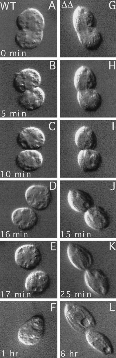
We cloned two genes, KIN1 and KIN2, encoding kinesin-II homologues from the ciliate Tetrahymena thermophila and constructed strains lacking either KIN1 or KIN2 or both genes. Cells with a single disruption of either gene showed partly overlapping sets of defects in cell growth, motility, ciliary assembly, and thermoresistance. Deletion of both genes resulted in loss of cilia and arrests in cytokinesis. Mutant cells were unable to assemble new cilia or to maintain preexisting cilia. Double knockout cells were not viable on a standard medium but could be grown on a modified medium on which growth does not depend on phagocytosis. Double knockout cells could be rescued by transformation with a gene encoding an epitope-tagged Kin1p. In growing cells, epitope-tagged Kin1p preferentially accumulated in cilia undergoing active assembly. Kin1p was also detected in the cell body but did not show any association with the cleavage furrow. The cell division arrests observed in kinesin-II knockout cells appear to be induced by the loss of cilia and resulting cell paralysis.


Figures

Figure 1
Figure 1
KIN1 and KIN2 are kinesin-II homologous genes in Tetrahymena. (A) Diagram of the alignment of predicted KIN1 and KIN2 encoded protein sequences. Domains are indicated as follows: stripes, motor domain; gray, coiled coil stalk; white, globular tail. Percent identity between Kin1p and Kin2p in each domain is indicated. Triangles represent positions of introns in the corresponding genomic DNA. (B) Phylogram of kinesin-II proteins. Alignments of multiple sequences were prepared using PILEUP; evolutionary distances between sequences were calculated using DISTANCES; and an evolutionary tree was made using GROWTREE of the UWGCG system (Devereux et al., 1984). (C) Sequence comparison between KIN1 and KIN2 and between KIN2 and Chlamydomonas reinhardtii FLA10 (Walther et al., 1994). Dot matrix analysis was done using COMPARE and DOTPLOT programs of UWGCG. Sequence data used for comparisons are available from European Molecular Biology Laboratory, GenBank, and DNA Data Bank of Japan under accession numbers AJ244020 (KIN1), AJ244021 (KIN2), L33697 (Crfla10), D14968 (Ceosm3), AB002357 (Hskiaa0359), A57107 (Mmkif3b), C48835 (Xlklp3), AF013116 (Mmkif3c), U00996 (Spkrp95), U15974 (Dmklp68d), D12645 (Mmkif3a), and L16993 (Spkrp85). The sequence of Ttkin5 was provided by M. Bernstein (personal communication). The Cekrp85 and Cekrp95 sequences were identified by the C. elegans sequencing project (Signor et al., 1999).
Figure 2
Figure 2
Germ line disruption of KIN1 and KIN2. (A). Genetic scheme for creation of homozygous single knockout strains. (1 and 2) During mating between two wild-type strains the targeted gene is disrupted in the MIC by biolistic transformation. The resulting progeny are heterozygous for the null allele in both the MAC and the MIC. (3) During vegetative growth heterozygous clones become homozygous for the wild-type allele in the MAC because of random segregation of alleles in the amitotic MAC (phenotypic assortment). (4–6) A strain heterozygous for the disrupted allele in the MIC is crossed to a star strain lacking a functional MIC, resulting in the transfer of a haploid MIC containing only a disrupted allele of the target gene. Endoreplication produces a diploid MIC yielding knockout heterokaryon cells. (7 and 8) Conjugation of two knockout heterokaryons leads to formation of a new MAC containing only disrupted copies of the targeted gene. (B) Analysis of germ line KIN1::neo2 transformants. Left panel, Southern blot of total genomic DNAs digested with EcoRI and BglII and probed with a radiolabeled KIN1 fragment. Right panel, diagram of the KIN1 locus. Lane 1, control strain DNA; lanes 2 and 3, independent germline transformants. The endogenous locus gives two fragments of 2.8 and 4 kb. A gene knockout is expected to give a single 8.2-kb fragment, and a 3′ integration should give 2.8- and 8.9-kb fragments. Note that the restriction patterns of transformant DNA analyzed in lane 3 is consistent with gene replacement, whereas in the transformant DNA analyzed in lane 2 the KIN1::neo2 fragment integrated into the 3′ flanking region of KIN1 gene. The endogenous 4.0-kb fragment is found in the original germ line transformants, indicating that these clones are heterozygous for KIN1/kin1::neo2. (C) Left panel, Southern blot of total genomic DNAs digested with Csp45 I and probed with a radiolabeled KIN2 genomic fragment. Right panel, diagram of the KIN2 locus. Lane 1, a transformant strain homozygous for KIN2::bsr1 gene; lane 2, wild-type control. The endogenous KIN2 gives a fragment of 3.0 kb. Disrupted KIN2 gives a fragment of 3.7 kb. The minor upper band present in both lanes most likely represents an incompletely digested KIN2 fragment.
Figure 3
Figure 3
Cytological analysis of ΔKIN1ΔKIN2. The phenotype was brought to expression by mating of double knockout heterokaryon strains (UG13 and UG14). Cells shown in A–H were grown in SPP medium, whereas the cell shown in I was grown in MEPP medium. Individual conjugation progeny cells were isolated and prepared for immunofluorescent confocal microscopy by staining with anti-tubulin antibodies (SG) and DAPI. (A) Dividing WT control 12 h after pair isolation. (B) WT control (84 h). (C) ΔKIN1ΔKIN2 cell (12 h) undergoing an early stage of cell division. The oral apparatus is already duplicated. (D) ΔKIN1ΔKIN2 cell (22 h) showing cilia heterogeneous in length. (E) ΔKIN1ΔKIN2 cell (29 h) at a final stage of cytokinesis. (F) ΔKIN1ΔKIN2 cell (29 h) with unseparated daughter cells and two sets of nuclei. (G and H) ΔKIN1ΔKIN2 cells (41 and 60 h) with uniformly short cilia, multiple nuclei, and multiple cortical subcells. (I) ΔKIN1ΔKIN2 cell 15 d after isolation of pairs grown in MEPP medium. Bars, 25 μm (bar in A shows scale for A–H). OA, oral apparatus.
Figure 4
Figure 4
(A–D) Electron microscopic analysis of WT and ΔKIN1ΔKIN2 cells. (A) Longitudinal section through WT axoneme. (B) Longitudinal section through axonemal remnant of a ΔKIN1ΔKIN2 cell. (C and D) Cross-sections through WT and double knockout basal bodies, respectively. Cells were processed for thin-section electron microscopy 84 h after pair isolation. (E) Analysis of the length of ciliary axonemes in ΔKIN1ΔKIN2 and WT cells, measured on confocal sections of cells labeled for tubulin by the SG serum using the NIH Image software package. Values are mean ± SD (n = 110 and 120 for wild-type and ΔKIN1ΔKIN2 cells, respectively).
Figure 5
Figure 5
Quantitative analysis of cell morphology and subunit composition. ΔKIN1ΔKIN2 and WT cells were prepared by mating of appropriate parental strains and isolation of individual mating pairs into either SPP or MEPP medium. Cells prepared for immunofluorescence microscopy were scored for the number of cortical subcells (A), number of MACs (B), and number of MICs (C). Wild-type and ΔKIN1ΔKIN2 cells grown in SPP were scored 60 h after isolation of pairs. ΔKIN1ΔKIN2 cells grown in MEPP were scored 15 d after pair isolation. Histograms show percentages of cells with the indicated numbers of cortical subunits or nuclei (n = 135). (D) Compiled data from observations on living cells and parallel immunofluorescence studies. Paralysis is measured as the percentage of drops containing paralyzed cells (n = 41), and cytokinesis failures are estimated as the percentage of cells prepared as in A–C containing multiple subunits (n = 10–135).
Figure 6
Figure 6
(A) Double knockout heterokaryon strains (UG13 and UG14) were crossed to express the ΔKIN1ΔKIN2 phenotype and rescued with gene fragments of either KIN1(−) or KIN1 containing an N-terminal 5xMyc epitope tag (+). Cells were selected with mp on SPP medium, and surviving, motile clones were analyzed by Western blotting of fractions prepared from cells rescued with either construct. The blot was probed with a monoclonal anti-myc antibody. A band of appropriate size (∼100 kDa) for 5xMyc-Kin1p is present only in ciliary fractions of cells transformed with epitope-tagged KIN1. (B and C) To verify the effectiveness of cell fractionation procedures, blots were probed with antibodies that recognize the macronuclear histone hv1 (B) or the AXO49 antibodies (C) directed against hyperglycylated tubulin isoforms, which are restricted to cilia (Bre et al., 1996).
Figure 7
Figure 7
Wild-type (A–F, M, and N) and GFP-kin1p rescued (G–L, O, and P) cells were isolated from exponentially growing cultures and processed for confocal analysis using anti-GFP antibodies and TAP952 anti-tubulin antibodies. GFP and tubulin were detected using secondary antibodies coupled to Cy3 and FITC, respectively. For individual cells corresponding images of GFP (A, C, E, G, I, K, M, and O) and tubulin (B, D, F, H, J, L, N, and P) are shown. (A and B) Negative control cell. Some background staining with GFP antibodies is present in the cell body (A). (C–F) higher magnifications of the boxes shown in panels A and B. (G and H) Nondividing cell expressing GFP-Kin1p. Note weak staining of cilia by anti-GFP antibodies. Some cilia shown in boxed areas are labeled more strongly by anti-GFP antibodies (G). These cilia are shorter and label more uniformly by the TAP952 antibodies (H). (I–L) Higher magnifications of the boxes shown in G and H. Arrowheads show shorter immature cilia, which label uniformly with TAP952 antibodies and also show more of GFP signal. Stars indicate mature cilia in which the TAP952 labeling is restricted to ciliary tips, and there is relatively less GFP signal. (M and N) Negative control dividing cell. Two oral apparatuses are present. (O and P) Dividing cell expressing GFP-Kin1p. The oral cilia are labeled heavily by anti-GFP antibodies (O) and uniformly by TAP952 antibodies (P). Note relatively weak staining for GFP in the oral apparatus of a nondividing cell (G). Bars: A and M, 15 μm; C, 1 μm. OA, oral apparatus; LM, longitudinal microtubule bundle.
Figure 8
Figure 8
Cells expressing GFP-Kin1p were starved for 24 h, deciliated, and allowed to regenerate cilia. At various times, samples of cells were processed for immunofluorescence using anti-GFP antibodies plus secondary antibodies coupled to Cy3 and TAP952 anti-tubulin antibodies plus secondary antibodies coupled to FITC. Pairs of corresponding GFP (A, C, E, and G) and tubulin (B, D, F, and H) images are shown for individual cells. Bars, 15 μm.
Figure 9
Figure 9
Comparison of cleavage furrow progression in WT (A–F) and ΔKIN1ΔKIN2 (G–L) cells. Single cells were isolated and analyzed using differential interference contrast video microscopy.

References

    1. Altschul SF, Gish W, Miller W, Myers EW, Lipman DJ. Sequence homology searches done using BLAST. J Mol Biol. 1990;215:403–410. - PubMed
    1. Bre M-H, et al. Axonemal tubulin polyglycylation probed with two monoclonal antibodies: widespread evolutionary distribution, appearance during spermatozoan maturation and possible function in motility. J Cell Sci. 1996;109:727–738. - PubMed
    1. Callen A-M, et al. Isolation and characterization of libraries of monoclonal antibodies directed against various forms of tubulin in Paramecium. Biol Cell. 1994;81:95–119. - PubMed
    1. Calzone FJ, Gorovsky MA. Cilia regeneration in Tetrahymena. Exp Cell Res. 1982;140:474–476. - PubMed
    1. Cassidy-Hanley D, Bowen J, Lee J, Cole ES, VerPlank LA, Gaertig J, Gorovsky MA, Bruns PJ. Germline and somatic transformation of mating Tetrahymena thermophila by particle bombardment. Genetics. 1997;146:135–147. - PMC - PubMed

Publication types

LinkOut - more resources