. 1999 Oct;10(10):3113-23.
doi: 10.1091/mbc.10.10.3113.

Testosterone signaling through internalizable surface receptors in androgen receptor-free macrophages

Affiliations
Free PMC article

Testosterone signaling through internalizable surface receptors in androgen receptor-free macrophages

W P Benten et al. Mol Biol Cell. 1999 Oct.
Free PMC article

Abstract

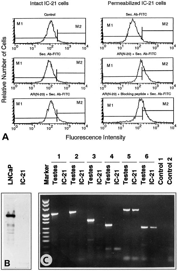
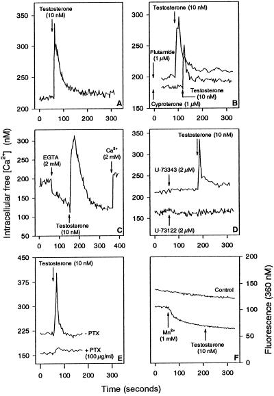
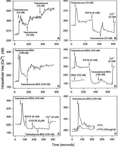
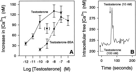
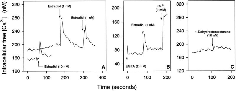
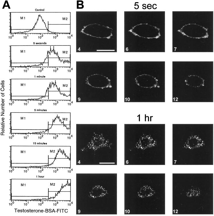
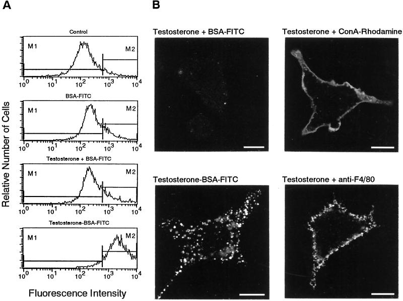
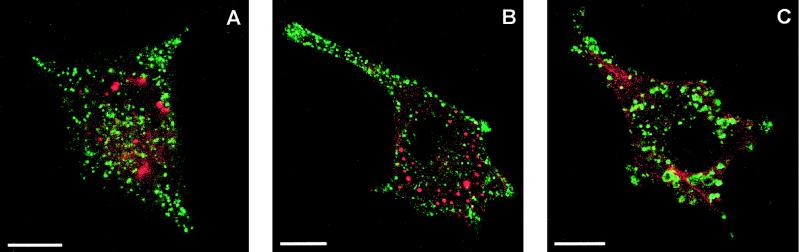
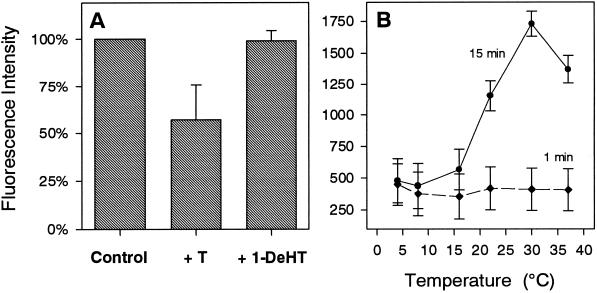
Testosterone acts on cells through intracellular transcription-regulating androgen receptors (ARs). Here, we show that mouse IC-21 macrophages lack the classical AR yet exhibit specific nongenomic responses to testosterone. These manifest themselves as testosterone-induced rapid increase in intracellular free [Ca(2+)], which is due to release of Ca(2+) from intracellular Ca(2+) stores. This Ca(2+) mobilization is also inducible by plasma membrane-impermeable testosterone-BSA. It is not affected by the AR blockers cyproterone and flutamide, whereas it is completely inhibited by the phospholipase C inhibitor U-73122 and pertussis toxin. Binding sites for testosterone are detectable on the surface of intact IC-21 cells, which become selectively internalized independent on caveolae and clathrin-coated vesicles upon agonist stimulation. Internalization is dependent on temperature, ATP, cytoskeletal elements, phospholipase C, and G-proteins. Collectively, our data provide evidence for the existence of G-protein-coupled, agonist-sequestrable receptors for testosterone in plasma membranes, which initiate a transcription-independent signaling pathway of testosterone.


Figures

Figure 1
Figure 1
Absence of intracellular AR in IC-21 cells. (A) Flow cytometry of intact and permeabilized IC-21 cells incubated for 1 h with the anti-AR antibody AR (N-20) and secondary fluorescent antibody (Sec. Ab-FITC). In permeabilized cells, the blocking peptide AR (N-20)P cannot competitively displace the slight increase in fluorescence of AR (N-20). (B) IC-21 cells and LNCaP cells as a control were subjected to Western blotting using the anti-AR antibody AR (N-20) and the ECL detection system. Only LNCaP cells reveal the AR band at 110 kDa. (C) RT-PCR with RNA isolated from mouse testes and IC-21 cells with markers (pUC mix, MBI Fermentas) on the left. The primer pair AR-P1/AR-M1 (1) spanned a 511-nt region of the DNA-binding domain of AR. The primer pairs AR-P2/AR-M2 (2), AR-P3/AR-M3 (3), and AR-P4/AR-M3 (4) spanned regions of the carboxyl terminus of the AR with 560, 365, and 281 nt, respectively. The expected bands were only revealed in testes. The primer pairs mzfm-P1/mzfm-M1 (5) and mzfm-P2/mzfm-M2 (6) yielded bands of 640 and 253 nt, respectively, of the low abundant mRNA of the gene mzfm in both testes and IC-21 cells. Control 1, RT-PCR without primers; Control 2, PCR with the primer pair AR-P1/AR-M1 without RNA.
Figure 2
Figure 2
Testosterone-induced Ca2+ mobilization in IC-21 cells. (A) Testosterone causes an immediate Ca2+ spike. (B) Cells were treated with cyproterone for 30 min or flutamide for 60 min before adding testosterone. (C) Cells were incubated with EGTA for 90 s before addition of testosterone. (D) Cells were treated with the phospholipase C inhibitor U-73122 or with the inactive control compound U-73343 for 2 min before adding testosterone. (E) Cells were pretreated with pertussis toxin for 16 h (+ PTX) before addition of testosterone. (F) Cells were incubated with Mn2+ for 2 min before adding testosterone. Arrows indicate addition of substances to IC-21 cells.
Figure 3
Figure 3
Ca2+ Responses of IC-21 cells to testosterone and testosterone-BSA. (A) A second or third addition of testosterone shortly after the first treatment induces a sustained increase in [Ca2+]i rather than a Ca2+ spike. (B) Cells were treated for 4 min with testosterone and then for 2 min with EGTA before the second addition of testosterone that induced only a Ca2+ spike. (C) The first addition of testosterone-BSA induced a Ca2+ spike, whereas the second addition induced a sustained Ca2+ increase. (D) BSA alone had no effect on [Ca2+]i (lower line). After incubation with testosterone-BSA for 2 min, cells were treated with EGTA before the second addition of testosterone-BSA. (E) After incubation of cells with testosterone-BSA, the cells were pretreated with both EGTA and U-73122 before the second addition of testosterone-BSA. (F) Cells were treated with pertussis toxin for 16 h (+ PTX) before adding testosterone-BSA. Arrows indicate addition of substances to IC-21 cells.
Figure 4
Figure 4
Effect of different concentrations of testosterone, testosterone-BSA, and testosterone pretreatment on [Ca2+]i of IC-21 cells. (A) Increase in [Ca2+]i with increasing concentrations of testosterone and testosterone-BSA, respectively. (B) Cells were pretreated with testosterone for 60 min before adding testosterone for a second time.
Figure 5
Figure 5
Effect of estradiol and 1-dehydrotestosterone on [Ca2+]i of IC-21 cells. (A) Treatment of cells with 1 nM estradiol resulted in a higher increase in [Ca2+]i than 10 nM estradiol. A second addition of estradiol shortly after the first addition again induced a Ca2+ spike. (B) Cells were incubated with EGTA for 1 min before adding estradiol. (C) Addition of 1-dehydrotestosterone had no effect on [Ca2+]i. Arrows indicate the addition of substances to IC-21 cells.
Figure 6
Figure 6
Surface binding sites of testosterone and their internalization in intact IC-21 cells. (A) Cells were incubated with testosterone-BSA-FITC for various periods between 5 s and 1 h and then analyzed by flow cytometry. (B) CLSM of cells incubated with testosterone-BSA-FITC for 5 s and 1 h. Optical slices of 0.5 μm. Bars, 10 μm.
Figure 7
Figure 7
Selective internalization of surface binding sites of testosterone. (A) Flow cytometry of IC-21 cells treated with the indicated substances for 15 min. (B) CLSM of cells incubated for 15 min with the indicated substances and the corresponding FITC-labeled secondary antibody. Note the difference between the smooth uniform surface labeling with Con A-rhodamine and the granular surface fluorescence of the F4/80 antigens. Bars, 10 μm.
Figure 8
Figure 8
CLSM colocalization of the sequestered surface binding sites of testosterone. (A) IC-21 cells were incubated in parallel with testosterone-BSA-FITC and LysoTracker Red DND-99 at 37°C for 1 h. Testosterone-BSA-FITC did not colocalize with acidic vesicles stained with LysoTracker Red DND-99. (B) Testosterone-BSA-FITC was not sequestrated within clathrin-coated vesicles as detected by anti-clathrin antibodies and the Cy3-labeled corresponding secondary antibody. (C) Internalized punctate fluorescence of testosterone-BSA-FITC was not labeled with an anti-caveolin antibody and its corresponding secondary TRITC-conjugated antibody. Bars, 10 μm.
Figure 9
Figure 9
Flow cytometry of the internalization of surface-bound testosterone-BSA-FITC in IC-21 cells. (A) Cells were incubated for 15 min with testosterone-BSA-FITC (10−6 M) in the absence (Control) or in the presence of a 10-fold excess of unlabeled testosterone (+ T) or 1-dehydrotestosterone (+ 1-DeHT). Values normalized to controls are given as means ± SD from five different experiments. (B) Cells were equilibrated for 30 min at the indicated temperatures and then incubated with 1.5 × 10⁻⁵ M testosterone-BSA-FITC for 1 or 15 min at the same temperatures. Values represent means ± SD from at least two different experiments.

References

    1. Beato M. Gene regulation by steroid hormones. Cell. 1989;56:335–344. - PubMed
    1. Benten WPM, Bettenhaeuser U, Wunderlich F, Van Vliet E, Mossmann H. Testosterone-induced abrogation of self-healing of Plasmodium chabaudi malaria in B10 mice: mediation by spleen cells. Infect Immun. 1991;59:4486–4490. - PMC - PubMed
    1. Benten WPM, Lieberherr M, Giese G, Wrehlke C, Stamm O, Sekeris CE, Mossmann H, Wunderlich F. Functional testosterone receptors in plasma membranes of T cells. FASEB J. 1999;13:123–133. - PubMed
    1. Benten WPM, Lieberherr M, Giese G, Wunderlich F. Estradiol binding to cell surface raises cytosolic free calcium in T cells. FEBS Lett. 1998;422:349–353. - PubMed
    1. Benten WPM, Lieberherr M, Sekeris CE, Wunderlich F. Testosterone induced Ca2+ influx via nongenomic surface receptors in activated T cells. FEBS Lett. 1997;407:211–214. - PubMed

MeSH terms