. 1999 Oct;10(10):3357-72.
doi: 10.1091/mbc.10.10.3357.

GERp95, a membrane-associated protein that belongs to a family of proteins involved in stem cell differentiation

Affiliations
Free PMC article

GERp95, a membrane-associated protein that belongs to a family of proteins involved in stem cell differentiation

D E Cikaluk et al. Mol Biol Cell. 1999 Oct.
Free PMC article

Abstract

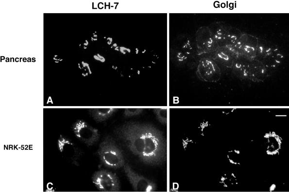
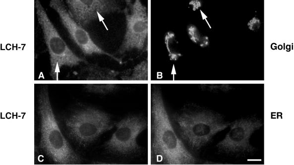
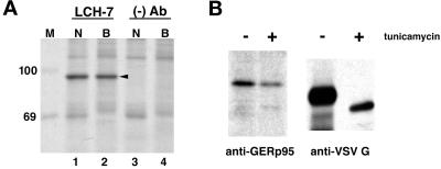
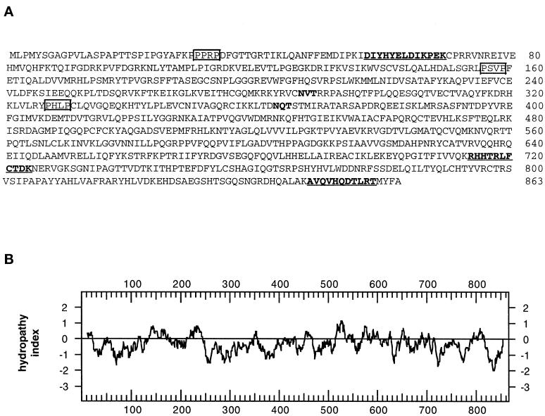
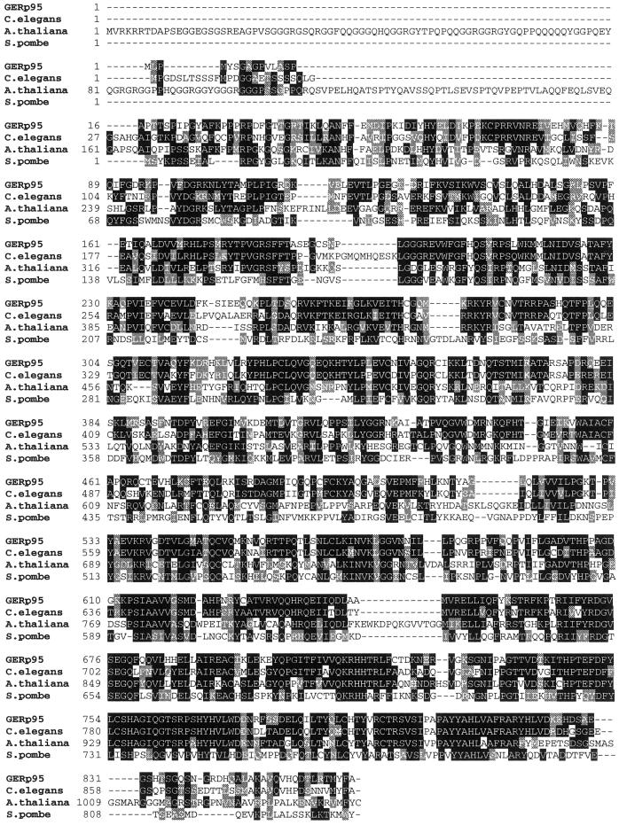
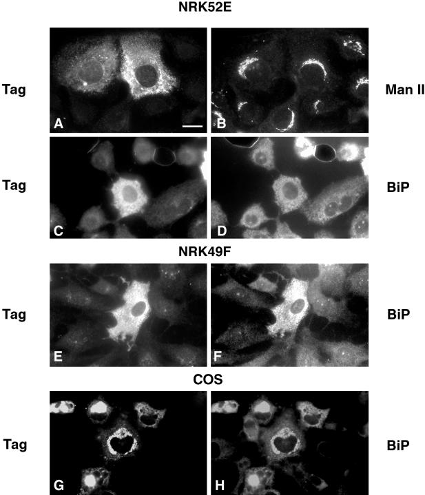
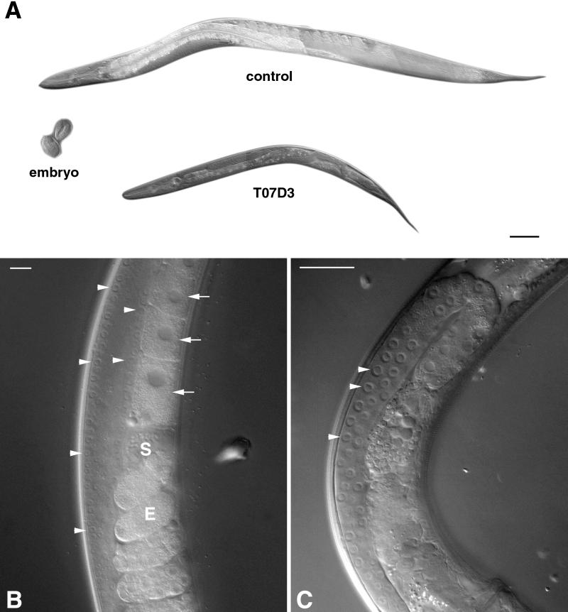
A panel of mAbs was elicited against intracellular membrane fractions from rat pancreas. One of the antibodies reacted with a 95-kDa protein that localizes primarily to the Golgi complex or the endoplasmic reticulum (ER), depending on cell type. The corresponding cDNA was cloned and sequenced and found to encode a protein of 97.6 kDa that we call GERp95 (Golgi ER protein 95 kDa). The protein copurifies with intracellular membranes but does not contain hydrophobic regions that could function as signal peptides or transmembrane domains. Biochemical analysis suggests that GERp95 is a cytoplasmically exposed peripheral membrane protein that exists in a protease-resistant complex. GERp95 belongs to a family of highly conserved proteins in metazoans and Schizosaccharomyces pombe. It has recently been determined that plant and Drosophila homologues of GERp95 are important for controlling the differentiation of stem cells (Bohmert et al., 1998; Cox et al., 1998; Moussian et al., 1998). In Caenorhabditis elegans, there are at least 20 members of this protein family. To this end, we have used RNA interference to show that the GERp95 orthologue in C. elegans is important for maturation of germ-line stem cells in the gonad. GERp95 and related proteins are an emerging new family of proteins that have important roles in metazoan development. The present study suggests that these proteins may exert their effects on cell differentiation from the level of intracellular membranes.


Figures

Figure 1
Figure 1
LCH-7 recognizes an antigen in the Golgi complex of pancreas acinar and epithelial NRK cells. Rat pancreas sections and NRK cells were fixed and processed for double-label indirect immunofluorescence as described. Samples were incubated with the mAb LCH-7 (A and C) and the Golgi marker rabbit anti-Man II (B and D). Colocalization of LCH-7 and Man II can be seen in the Golgi of pancreas acinar and NRK52E cells (A–D). A limited amount of staining for Man II is also observed on the plasma membrane of the acinar cells (B). Bar in D, ∼10 μm.
Figure 2
Figure 2
LCH-7 stains ER-associated structures in fibroblastic NRK cells. Cells were processed for double-label indirect immunofluorescence by staining with LCH-7 (A and C) and rabbit anti-Man II to label Golgi (B) or rabbit anti-ERp72 to label ER (D). No colocalization between LCH-7 and anti-Man II is evident (A and B, arrowheads), but some overlap is seen with the anti-ER antibody (C and D). Bar, ∼10 μm.
Figure 3
Figure 3
Immunoprecipiation of GERp95 from cultured cells. (A) NRK52E (N) and BHK-21 (B) cells were biosynthetically labeled with 35S cysteine/methionine for 6 h before lysis and immunoprecipitation with LCH-7 or a mAb to CD8 ((−) Ab). Samples were subjected to SDS-PAGE on 8% gels followed by fluorography. A 95-kDa protein, GERp95 (arrowhead), is immunoprecipitated from both cell types with the LCH-7 antibody (lanes 1 and 2) but not the negative control Ab (lanes 3 and 4). 14C-labeled protein standards (kDa) are shown lane M. (B) NRK52E cells were biosynthetically labeled with 35S as above in the presence and absence of tunicamycin (3 μg/ml) before lysis and immunoprecipitation with LCH-7. Cells were infected with VSV and radiolabeled using the same conditions, and the VSV G protein was immunoprecipitated and subjected to SDS-PAGE and fluorography. The mobility of VSV G is increased in the presence of tunicamycin attributable to inhibition of N-linked glycosylation.
Figure 4
Figure 4
Predicted amino acid sequence and hydropathy analysis of GERp95. (A) Predicted amino acid sequence of GERp95. Internal peptide sequences obtained by microsequencing immunoaffinity-purified protein are underlined and shown in bold. Two consensus sites for N-linked glycosylation are highlighted in bold. Potential high-affinity SH3-binding sites (PxxP) are boxed. (B) Hydropathy analysis of GERp95 according to the algorithm of Kyte and Doolittle (1982) using a window size of 19 amino acids for scanning. (C) Sequence alignment of rat GERp95 and related proteins from C. elegans, A. thaliana, and S. pombe. Identical and conserved amino acids are indicated by shading in black and gray, respectively. The genomes of C. elegans and A. thaliana each encode multiple GERp95-related proteins, but only the closest relatives are shown in this figure.
Figure 4
Figure 4
Predicted amino acid sequence and hydropathy analysis of GERp95. (A) Predicted amino acid sequence of GERp95. Internal peptide sequences obtained by microsequencing immunoaffinity-purified protein are underlined and shown in bold. Two consensus sites for N-linked glycosylation are highlighted in bold. Potential high-affinity SH3-binding sites (PxxP) are boxed. (B) Hydropathy analysis of GERp95 according to the algorithm of Kyte and Doolittle (1982) using a window size of 19 amino acids for scanning. (C) Sequence alignment of rat GERp95 and related proteins from C. elegans, A. thaliana, and S. pombe. Identical and conserved amino acids are indicated by shading in black and gray, respectively. The genomes of C. elegans and A. thaliana each encode multiple GERp95-related proteins, but only the closest relatives are shown in this figure.
Figure 5
Figure 5
Expression of GERp95-specific RNA in mouse tissues. A membrane containing 50 μg of total RNA isolated from mouse tissues was hybridized with a 32P-labeled cDNA probe derived from the 5′ coding region of the rat GERp95 cDNA. The blot was stripped and probed with a radiolabeled cyclophilin cDNA to show relative loading and integrity of RNA. The positions the 28S and 18S rRNAs are indicated.
Figure 6
Figure 6
Characterization of a polyclonal antibody to recombinant GERp95. (A) GERp95 was expressed in vitro using a coupled transcription/translation system containing 35S methionine. The samples were subjected to immunoprecipitation with LCH-7, rabbit preimmune, or anti-GERp95 sera. Samples were subjected to SDS-PAGE on 8% gels followed by fluorography. Both LCH-7 and rabbit anti-GERp95 precipitate a major band of ∼95 kDa. (B) NRK49F and NRK52E cells were biosynthetically labeled with 35S cysteine/methionine for 4 h before lysis and immunoprecipitation with rabbit preimmune or anti-GERp95 serum. Rabbit anti-GERp95 serum specifically immunoprecipitates a protein with an apparent molecular weight of ∼95 kDa from both NRK49F and NRK52E cells. (C) NRK52E lysates were separated by SDS-PAGE, transferred to PVDF membrane, and probed with rabbit preimmune or anti-GERp95 serum. (D) NRK52E (a and b) and NRK49F (c and d) cells fixed with acid-alcohol were incubated with LCH-7 and rabbit anti-GERp95 serum. Both LCH-7 and rabbit anti-GERp95 stain the Golgi complex in NRK52E cells (a and b) but not in NRK49F cells (c and d). Bar, 10 μm.
Figure 7
Figure 7
Interaction of GERp95 with membranes in vivo and in vitro. (A) Crude microsomes and cytosol were prepared from NRK52E and NRK49F cells, and equivalent proportions normalized to starting volumes were separated by SDS-PAGE and transferred to PVDF membranes. The membranes were probed with rabbit antisera to GERp95, calnexin, and rat anti-HSP70. The majority of GERp95 and calnexin are found in the membrane fractions, whereas HSP70 is in the soluble fraction. (B) Fifty micrograms (protein) of rat liver fractions were subjected to SDS-PAGE and immunoblotting with antibodies to GERp95, and calnexin (ER marker) and Man II (Golgi). The highest concentrations of GERp95 are found in rough ER fractions. (C) NRK52E microsomes were extracted with HME, 0.5 M KCl, 1.0 M KCl, or Hi pH buffer (0.1 M sodium carbonate, pH 11.5), and then centrifuged at 100,000 × g for 60 min to obtain pellet (P) and soluble (S) fractions. Alkaline treatment and KCl washing resulted in complete extraction of GERp95 from membranes. (D) Microsomes were incubated at 0°C with increasing amounts of trypsin/chymotrypsin with or without 1% Triton X-100 followed by SDS-PAGE and immunoblotting with rabbit antiserum to GERp95, β-COP, and glucosidase II β-subunit. In the absence of detergent, GERp95 and glucosidase II β are insensitive to protease, but are completely digested when Triton X-100 is included. In contrast, β-COP, a peripheral membrane protein on the cytosolic side of membranes is completely degraded by the proteases even in the absence of detergent. (E) 35S-labeled GERp95 was synthesized in vitro in the presence or absence of canine pancreatic microsomes. Samples were subjected to digestion with varying amounts of trypsin/chymotrypsin in the presence or absence of Triton X-100 before SDS-PAGE and autoradiography. VSV G protein was used as a positive control to show translocational activity of the microsomes. VSV G is only protected from protease when microsomes are present and detergent is absent.
Figure 8
Figure 8
Expression of liver-derived GERp95 cDNA in NRK and COS cells. Cells were infected with recombinant Sindbis viruses encoding liver-derived GERp95 cDNA, which includes an epitope tag. Sixteen hours after infection, cells were processed for double-label indirect immunofluorescence using rabbit anti-tag (A, C, E, and G), mouse anti-Man II (B), and mouse anti-BiP (D, F, and H). COS cells were transfected with a mammalian expression vector encoding epitope-tagged GERp95 and processed for indirect immunofluorescence 24–40 h later. Bar, 10 μm.
Figure 9
Figure 9
The GERp95 orthologue in C. elegans is important for oocyte development. Gonads of young hermaphrodite worms were injected with double-stranded RNA synthesized from empty pBluescript vector (control) or a C. elegans GERp95-like gene (T07D3) on chromosome II. A shows the progeny of injected worms that are now adult hermaphrodites of the same age. Two wild-type embryos are shown for comparison. Bar, ∼100 μm. Higher magnifications of the gonad regions of injected worms are shown in B and C. In control worms (B), germ-line stem cells in the mitotic proliferation zone of the distal gonad are indicated by arrowheads. Brick-shaped oocytes with large nuclei are indicated by arrows. Sperm (S) and eggs (E) are also visible in the proximal gonads of control worms but not in progeny of T07D3-injected animals (C). In the progeny of T07D3-injected worms, normal-looking germ-line stem cells are present in the distal gonad (arrowheads), but oocytes and sperm are absent from the proximal gonad. Bars in B and C, ∼25 μm.

References

    1. Altschul S, Gish W, Miller W, Myers E, Lipman D. Basic local alignment search tool. J Mol Biol. 1990;215:403–410. - PubMed
    1. Arduengo PM, Appleberry OK, Chuang P, L’Hernault SW. The presenilin protein family member SPE-4 localizes to an ER/Golgi derived organelle and is required for proper cytoplasmic partitioning during Caenorhabditis elegans spermatogenesis. J Cell Sci. 1998;111:3645–54. - PubMed
    1. Arendt CW, Ostergaard HL. Identification of the CD45-associated 116-kDa and 80-kDa proteins as the alpha- and beta-subunits of the alpha-glucosidase II. J Biol Chem. 1997;272:13117–13125. - PubMed
    1. Balch WE, Dunphy WG, Braell WA, Rothman JE. Reconstitution of the transport of protein between successive compartments of the Golgi measured by the coupled incorporation of N-acetylglucosamine. Cell. 1984;39:405–416. - PubMed
    1. Beck KA, Buchanan J, Malhotra V, Nelson WJ. Golgi spectrin: identification of an erythroid β-spectrin homolog associated with the Golgi complex. J Cell Biol. 1994;127:707–723. - PMC - PubMed

Publication types

MeSH terms

Associated data