

Analysis of genomic alterations in sporadic adrenocortical lesions. Gain of chromosome 17 is an early event in adrenocortical tumorigenesis
- PMID: 10514385
- PMCID: PMC1867020
- DOI: 10.1016/S0002-9440(10)65205-4
Analysis of genomic alterations in sporadic adrenocortical lesions. Gain of chromosome 17 is an early event in adrenocortical tumorigenesis
Abstract
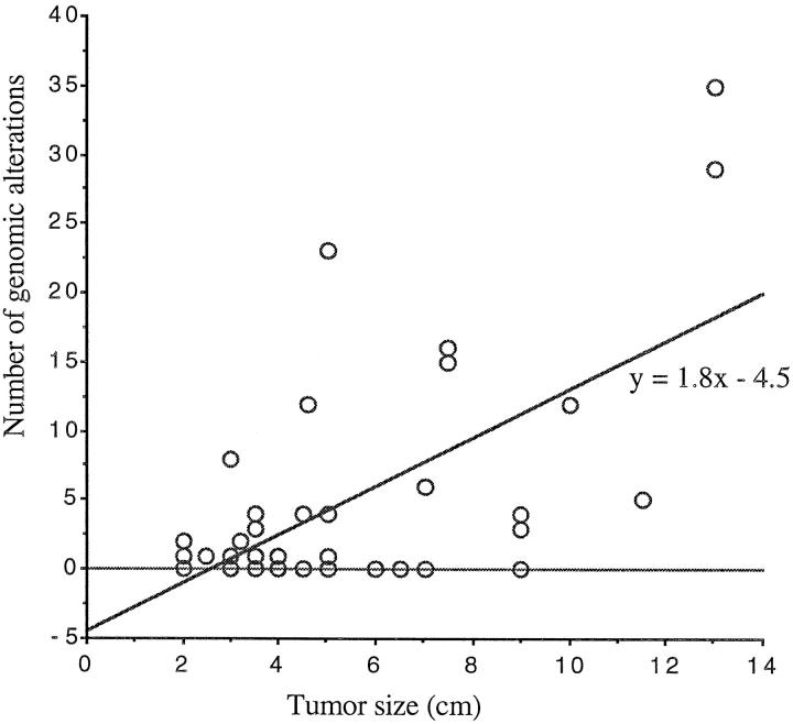
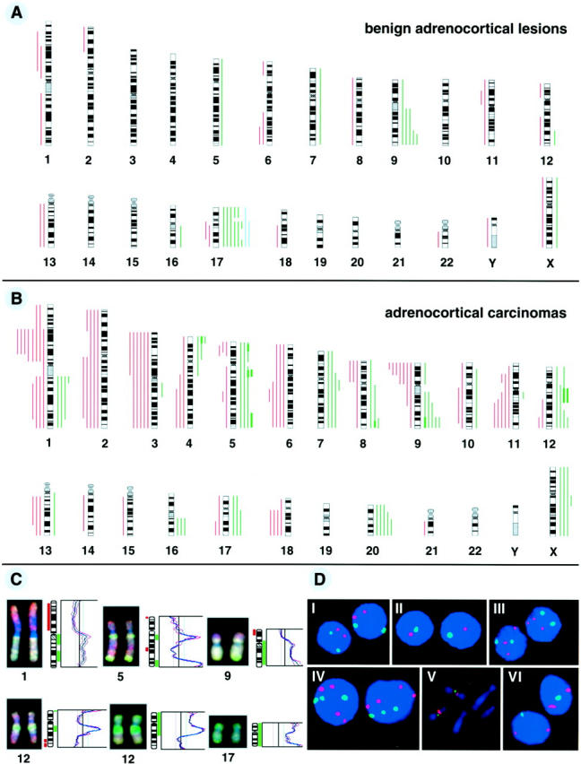
Genetic changes underlying the tumorigenesis of sporadic adrenocortical tumors are poorly characterized. To search for characteristic genomic imbalances involved in adrenocortical tumors, we examined 41 adrenocortical lesions (12 carcinomas, 23 adenomas, and 6 hyperplasias) by comparative genomic hybridization. Our results show that genetic alterations are more frequent in malignant than in benign lesions and that they rarely occur in hyperplastic lesions. The most frequent DNA copy number changes in adrenocortical carcinomas included losses of 1p21-31, 2q, 3p, 3q, 6q, 9p, and 11q14-qter, as well as gains and amplifications of 5q12, 9q32-qter, 12q, and 20q. The genomic aberrations prevalently occurring in adrenocortical adenomas were gains of 17q, 17p, and 9q32-qter. Gains found in 2 of 6 adrenocortical hyperplastic lesions involved chromosome 17 or 17q only. These data indicate that oncogenes determining the early tumorigenesis of adrenocortical tumors may exist on chromosome 17 and that the number of genomic alterations is closely associated with tumor behavior in adrenocortical tumors.
Figures
Similar articles
-
Adrenocortical carcinoma is characterized by a high frequency of chromosomal gains and high-level amplifications.Genes Chromosomes Cancer. 2000 Jun;28(2):145-52. Genes Chromosomes Cancer. 2000. PMID: 10824999
-
Genetic aberrations in adrenocortical tumors detected using comparative genomic hybridization correlate with tumor size and malignancy.Cancer Res. 1996 Sep 15;56(18):4219-23. Cancer Res. 1996. PMID: 8797595
-
Genomic aberrations in carcinomas of the uterine corpus.Genes Chromosomes Cancer. 2004 Jul;40(3):229-46. doi: 10.1002/gcc.20038. Genes Chromosomes Cancer. 2004. PMID: 15139002
-
Molecular markers and the pathogenesis of adrenocortical cancer.Oncologist. 2008 May;13(5):548-61. doi: 10.1634/theoncologist.2007-0243. Oncologist. 2008. PMID: 18515740 Review.
-
Gene expression profiling in adrenocortical neoplasia.Mol Cell Endocrinol. 2012 Mar 31;351(1):111-7. doi: 10.1016/j.mce.2011.09.044. Epub 2011 Oct 25. Mol Cell Endocrinol. 2012. PMID: 22056416 Review.
Cited by
-
GATA3 and APOBEC3B are prognostic markers in adrenocortical carcinoma and APOBEC3B is directly transcriptionally regulated by GATA3.Oncotarget. 2020 Sep 8;11(36):3354-3370. doi: 10.18632/oncotarget.27703. eCollection 2020 Sep 8. Oncotarget. 2020. PMID: 32934779 Free PMC article.
-
Towards an understanding of the role of p53 in adrenocortical carcinogenesis.Mol Cell Endocrinol. 2012 Mar 31;351(1):101-10. doi: 10.1016/j.mce.2011.09.010. Epub 2011 Sep 10. Mol Cell Endocrinol. 2012. PMID: 21930187 Free PMC article. Review.
-
Pediatric adrenocortical tumors: what they can tell us on adrenal development and comparison with adult adrenal tumors.Front Endocrinol (Lausanne). 2015 Feb 18;6:23. doi: 10.3389/fendo.2015.00023. eCollection 2015. Front Endocrinol (Lausanne). 2015. PMID: 25741319 Free PMC article. Review.
-
MicroRNA profiling of adrenocortical tumors reveals miR-483 as a marker of malignancy.Cancer. 2011 Apr 15;117(8):1630-9. doi: 10.1002/cncr.25724. Epub 2010 Nov 8. Cancer. 2011. PMID: 21472710 Free PMC article.
-
Improvement of histopathological classification of adrenal gland tumors by genetic differentiation.World J Urol. 2010 Jun;28(3):329-34. doi: 10.1007/s00345-010-0541-7. Epub 2010 Apr 3. World J Urol. 2010. PMID: 20364258
References
-
- Ross NS, Aron DC: Hormonal evaluation of the patient with an incidentally discovered adrenal mass. N Engl J Med 1990, 323:1401-1405 - PubMed
-
- Barzilay JI, Pazianos AG: Adrenocortical carcinoma. Urol Clin North Am 1989, 16:457-468 - PubMed
-
- Schröder S, Komminoth P, Padberg B, Heitz PU: Morphological typing, evaluation of tumor dignity and prognosis and etiologic classification of adrenomedullary and adrenocortical neoplasias. Pathologe 1995, 16:307-314 - PubMed
-
- Chandrasekharappa SC, Guru SC, Manickam P, Olufemi S, Collins FS, Emmert-Buck MR, Debelenko LV, Zhuang Z, Lubensky IA, Liotta LA, Crabtree JS, Wang Y, Roe BA, Weisemann J, Boguski MS, Agarwal SK, Kester MB, Kim YS, Heppner C, Dong Q, Spiegel AM, Burns AL, Marx SJ: Positional cloning of the gene for multiple endocrine neoplasia-type 1. Science 1997, 276:404-406 - PubMed
-
- Srivastava S, Zou ZQ, Pirollo K, Blattner W, Chang EH: Germ-line transmission of a mutated p53 gene in a cancer-prone family with Li-Fraumeni syndrome. Nature 1990, 348:747-749 - PubMed
Publication types
MeSH terms
LinkOut - more resources
Full Text Sources
