Clinical Trial
. 1999 Oct;155(4):1137-46.
doi: 10.1016/S0002-9440(10)65217-0.

Topical estrogen accelerates cutaneous wound healing in aged humans associated with an altered inflammatory response

Affiliations
Clinical Trial

Topical estrogen accelerates cutaneous wound healing in aged humans associated with an altered inflammatory response

G S Ashcroft et al. Am J Pathol. 1999 Oct.

Abstract

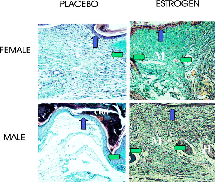
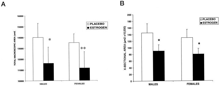
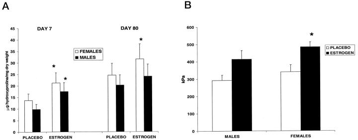
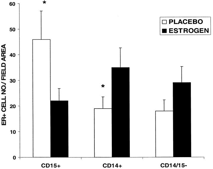
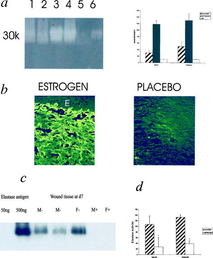
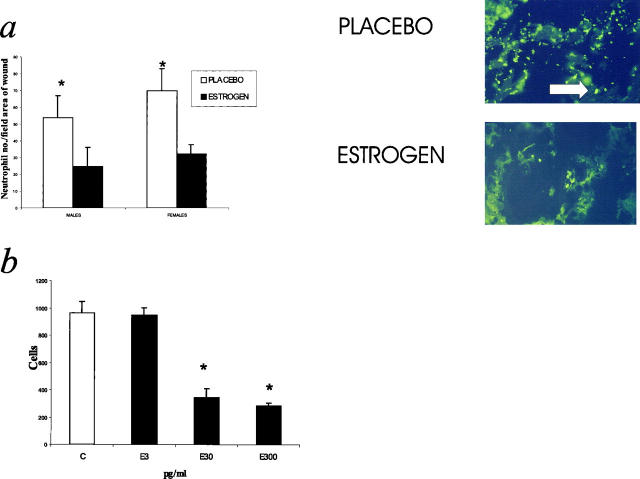
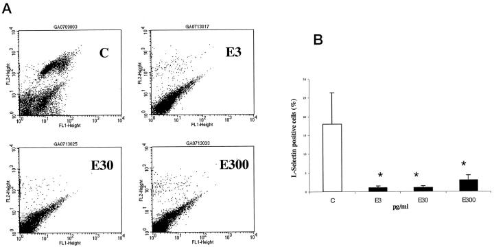
The effects of intrinsic aging on the cutaneous wound healing process are profound, and the resulting acute and chronic wound morbidity imposes a substantial burden on health services. We have investigated the effects of topical estrogen on cutaneous wound healing in healthy elderly men and women, and related these effects to the inflammatory response and local elastase levels, an enzyme known to be up-regulated in impaired wound healing states. Eighteen health status-defined females (mean age, 74.4 years) and eighteen males (mean age, 70.7 years) were randomized in a double-blind study to either active estrogen patch or identical placebo patch attached for 24 hours to the upper inner arm, through which two 4-mm punch biopsies were made. The wounds were excised at either day 7 or day 80 post-wounding. Compared to placebo, estrogen treatment increased the extent of wound healing in both males and females with a decrease in wound size at day 7, increased collagen levels at both days 7 and 80, and increased day 7 fibronectin levels. In addition, estrogen enhanced the strength of day 80 wounds. Estrogen treatment was associated with a decrease in wound elastase levels secondary to reduced neutrophil numbers, and decreased fibronectin degradation. In vitro studies using isolated human neutrophils indicate that one mechanism underlying the altered inflammatory response involves both a direct inhibition of neutrophil chemotaxis by estrogen and an altered expression of neutrophil adhesion molecules. These data demonstrate that delays in wound healing in the elderly can be significantly diminished by topical estrogen in both male and female subjects.


Figures

Figure 1.
Figure 1.
The deposition of matrix at day 7 was enhanced in elderly males and females by topical estrogen. Estrogen treatment stimulates matrix deposition (green fibers; M, matrix; H, hair follicle) in day 7 Masson’s Trichrome-stained sections. In placebo-treated wounds the granulation tissue is immature with fibrin deposits and little collagen. Horizontal arrows demarcate the wound edges (which, because of the increase in wound size, extend beyond the left field of view in the placebo-treated group). Vertical arrows indicate the epidermis. Original magnification, ×16.
Figure 2.
Figure 2.
Estrogen treatment was associated with reduced wound areas at day 7. A: Macroscopic areas as determined by planimetry (as described in Methods) were significantly reduced with estrogen treatment at day 7 post-wounding. *P = 0.04; **P = 0.02; n = 5 per group. B: Cross-sectional day 7 wound areas as assessed histologically using image analysis. Estrogen significantly reduced the wound cross-sectional area as determined using image analysis. *P < 0.05; n = 5 per group.
Figure 3.
Figure 3.
Estrogen increased wound collagen deposition and increased wound strength at day 80. A: Hydroxyproline levels were increased significantly in day 7 wound tissue by estrogen treatment for both males and females. At day 80 the difference between placebo and estrogen was only significant in females. *P < 0.05; n = 5 per group (d7) and n = 4 per group (d80). B: Mechanical properties of day 80 wounds assessed by the DAS showed an increase in wound stiffness at day 80 in the estrogen-treated wounds, which was significant only in females. *P < 0.05, n = 4 per group.
Figure 4.
Figure 4.
Estrogen receptor localization in acute wounds. Quantification of estrogen receptor-positive (ER+) cells in specific subgroups at day 7 post-wounding showed a significant increase in ER+CD15+ (neutrophil) cells and decrease in ER+ CD14+ (monocyte) cells in the placebo versus estrogen-treated wounds. *P < 0.05, n = 5 per group. ER+ CD14− CD15− cells were considered to be fibroblasts on morphological grounds and were found to be increased (not significantly) in the estrogen-treated wounds. No gender differences were observed. The data represented graphically are that of the male groups.
Figure 5.
Figure 5.
a: Factors capable of fibronectin breakdown in acute wounds are reduced with estrogen treatment. Fibronectin degradation is decreased in acute wounds of aged humans treated with estrogen compared to placebo. Degradation bands appear as pale regions in the gel. Lane 1, female-estrogen; 2, male-estrogen; 3, female-placebo; 4, male-placebo; 5, female skin; 6, 10 ng human neutrophil elastase. NS, normal skin. Band intensities were quantified using image analysis. Results are depicted as means ± SD. Each bar represents the average of band intensities from n = 5 subjects and thus the gel is a general representation. b: Fibronectin immunostaining is markedly increased at day 7 in estrogen-treated wounds. A marked increase in immunostaining for fibronectin was observed in the wounds of the estrogen-treated subjects for both males and females, compared to placebo. E, epidermis. In b no epithelium was present over the wound but photograph of section was taken below clot. Original magnification, ×25. c: Protease responsible for fibronectin degradation confirmed as elastase in acute wound tissue. Immunoblotting with an anti-human neutrophil elastase antibody confirmed that day 7 healing wounds in placebo-treated groups contained elastase, which localized to the degradation band seen in a parallel zymogram. Experiment shown is representative of n = 8 males, n = 8 females. Tissue from placebo groups contained >50 ng elastase/20 μg dry weight, whereas tissue from estrogen-treated groups contained <50 ng/20 μg dry weight. The sensitivity of this assay was >80 ng elastase, whereas the zymography is known to be a more sensitive assay for protease detection (in Figure 4A ▶ 10 ng elastase can be detected). M, males; F, females; +, estrogen; −, placebo. d: Elastase activity in acute wound tissue is reduced with estrogen treatment. Elastase activity was quantified using a synthetic elastase substrate degradation assay. *P < 0.05; n = 5 for each group. Elastase activity is expressed as ng/ml activity per 20 μg dry weight.
Figure 6.
Figure 6.
Neutrophil numbers were reduced in day 7 estrogen-treated wounds. a: Quantification of neutrophils in day 7 wounds. Estrogen treatment significantly reduced the numbers of local neutrophils in day 7 wounds *P < 0.05; n = 5 for each group. The immunostaining panels represent the center of day 7 wounds stained with an antibody to CD15. Original magnification, ×25. Arrows indicate CD15+ cells. b: Estrogen directly inhibited neutrophil chemotaxis. Estrogen treatment of neutrophils inhibited chemotaxis to fMLP at 30 and 300 pg/ml. n = 8. C, control.
Figure 7.
Figure 7.
Estrogen reduces the subpopulation of neutrophils expressing L-selectin. A: Representative FACS analysis of single-labeled neutrophils with L-selectin antibody. C, control; E3, estrogen 3 pg/ml, E30, estrogen 30 pg/ml; E300, estrogen 300 pg/ml. L-selectin cell population observed in the upper right quadrant at 10 (FL2) in control panel, and is largely absent after estrogen treatment for 12 hours. B: Reduction of L-selectin cell surface expression detected by FACS after estrogen treatment. n = 8. C, control. Gender had no effect on either parameter when male and female responses were assessed independently. *P < 0.05.

References

    1. Ashcroft GS, Horan MA, Ferguson MWJ: A review: The effects of ageing on cutaneous wound healing in mammals. J Anat 1995, 87:1-27 - PMC - PubMed
    1. Ashcroft GS, Horan MA, Herrick SE, Tarnuzzer RW, Schultz GS, Ferguson MWJ: Age-related differences in the temporal and spatial regulation of Matrix Metalloproteinases (MMPs) in normal skin and acute cutaneous wounds of healthy humans. Cell Tissue Res 1997, 290:581-591 - PubMed
    1. Herrick SE, Ashcroft GS, Ireland G, Horan MA, McCollum C, Ferguson MWJ: Up-regulation of elastase in acute wounds of healthy aged humans and chronic venous leg ulcers is associated with matrix degradation. Lab Invest 1996, 77:281-288 - PubMed
    1. McDonald JA, Kelley DG: Degradation of fibronectin by human leucocyte elastase. J Biol Chem 1980, 255:8848-8858 - PubMed
    1. Kafienah W, Buttle DJ, Burnett D, Hollander AP: Cleavage of type I collagen by human neutrophil elastase. Biochem J 1998, 330:897-902 - PMC - PubMed

Publication types

MeSH terms

LinkOut - more resources