. 1999 Oct;155(4):1173-81.
doi: 10.1016/s0002-9440(10)65220-0.

Non-Abeta component of Alzheimer's disease amyloid (NAC) revisited. NAC and alpha-synuclein are not associated with Abeta amyloid

Affiliations

Non-Abeta component of Alzheimer's disease amyloid (NAC) revisited. NAC and alpha-synuclein are not associated with Abeta amyloid

J G Culvenor et al. Am J Pathol. 1999 Oct.

Abstract

alpha-Synuclein (alphaSN), also termed the precursor of the non-Abeta component of Alzheimer's disease (AD) amyloid (NACP), is a major component of Lewy bodies and Lewy neurites pathognomonic of Parkinson's disease (PD) and dementia with Lewy bodies (DLB). A fragment of alphaSN termed the non-Abeta component of AD amyloid (NAC) had previously been identified as a constituent of AD amyloid plaques. To clarify the relationship of NAC and alphaSN with Abeta plaques, antibodies were raised to three domains of alphaSN. All antibodies produced punctate labeling of human cortex and strong labeling of Lewy bodies. Using antibodies to alphaSN(75-91) to label cortical and hippocampal sections of pathologically proven AD cases, we found no evidence for NAC in Abeta amyloid plaques. Double labeling of tissue sections in mixed DLB/AD cases revealed alphaSN in dystrophic neuritic processes, some of which were in close association with Abeta plaques restricted to the CA1 hippocampal region. In brain homogenates alphaSN was predominantly recovered in the cytosolic fraction as a 16-kd protein on Western analysis; however, significant amounts of aggregated and alphaSN fragments were also found in urea extracts of SDS-insoluble material from DLB and PD cases. NAC antibodies identified an endogenous fragment of 6 kd in the cytosolic and urea-soluble brain fractions. This fragment may be produced as a consequence of alphaSN aggregation or alternatively may accelerate aggregation of the full-length alphaSN.


Figures

Figure 1.
Figure 1.
αSN immunoreactivity of neuronal inclusions in DLB brain tissue. A: Labeled Lewy body in cingulate gyrus cortex using N-terminal Ab 97/5. B: Labeled Lewy bodies and neurite in substantia nigra using NAC Ab 42580. C: Labeled Lewy body in cingulate gyrus cortex using C-terminal domain Ab 97/8. Scale bars, 20 μm.
Figure 2.
Figure 2.
Western blot analysis of synuclein antibody reactivity on human brain homogenate from the cingulate gyrus cortex from DLB case. 10 μg of protein per lane was analyzed on 10% Tris-tricine gel. Antibodies were preabsorbed with peptide as indicated. A: Ab 97/5 at 1:5000 dilution. B: Ab 42580 at 1:1000 dilution. C: Ab 97/8 at 1:20,000. D: Ab 98/13 at 1:1000.
Figure 3.
Figure 3.
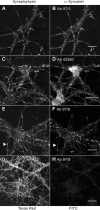
Confocal laser micrographs of double labeling of rat neurons stained for synaptophysin (Texas Red) and αSN (FITC). A, B: Labeling for synaptophysin showed almost complete co-localization with Ab 97/5 diluted 1:1000 after formaldehyde fixation. C, D: Labeling with NAC Ab 42580 at 1:100 after formaldehyde fixation also showed a punctate labeling with a higher apparent background over neuronal cell bodies, and there was considerable overlap with synaptophysin labeling. E, F: Labeling for synaptophysin showed considerable co-localization with Ab 97/8 diluted 1:1000 after formaldehyde fixation; some synaptophysin-positive punctate staining (arrowheads) did not have corresponding synuclein staining. G, H: Labeling after acetone fixation alone showed strong synaptophysin labeling but only very weak synuclein labeling (H). Scale bars, 20 μm.
Figure 4.
Figure 4.
Comparison Aβ and NAC antibody immunoreactivities in amyloid plaques in AD brain sections. A: AD frontal cortex stained with Aβ MAb 1E8 shows numerous plaques. B: A serial section from the same region as used in A stained in parallel with NAC Ab 42580 showed no NAC plaque labeling. C: AD hippocampus CA2 region stained with Aβ MAb 1E8 revealed numerous Aβ-positive plaques. D: The same region as used in C showed no plaque labeling with NAC Ab 42580. Scale bars, 50 μm.
Figure 5.
Figure 5.
Double labeling of CA1 region of hippocampus sections from DLB/AD case. A, B: αSN labeling (Ab 97/8) in brown and Aβ labeling (MAb 1E8) in blue shows αSN-positive globular neuritic structures surrounding Aβ-positive plaques (arrowheads in A), and αSN-positive globular clusters separate from a nearby Aβ-positive plaque. C, D: αSN labeling (Ab 97/8) in brown and tau labeling in red shows αSN-positive inclusions in neuron with tau-positive intraneuronal staining (arrows in C) and close association of globular αSN-positive neuritic processes with tau-positive structures (in D). Scale bars, 50 μm.
Figure 6.
Figure 6.
Western blot analysis of αSN expression in human brain fractions extracted from cingulate gyrus cortex of 2 cases each of DLB, PD, control, and AD. 5 μg of protein TBS-soluble, 10 μg of protein SDS-soluble, and 30 μl of urea-soluble fractions were applied per lane and run on separate gels. Due to slight gel variation separate markers were used in each gel as a reference. Blots were incubated with Ab 97/8 at 1:20,000 and exposed to ECL film for 1 minute, Ab 42580 at 1:2000 and exposed to ECL film for 5 minutes, or MAb WO2 at 1 μg/ml and exposed to film 1 minute (SDS) and 5 minutes (TBS).

Comment in

Similar articles

Cited by

References

    1. Krüger R, Kuhn W, Müller T, Woitalla D, Graeber M, Kösel S, Przuntek H, Epplen JT, Schöls L, Riess O: Ala30Pro mutation in the gene encoding α-synuclein in Parkinson’s disease. Nat Genet 1998, 18:106-108 - PubMed
    1. Polymeropoulos MH, Lavedan C, Leroy E, Ide SE, Dehejia A, Dutra A, Pike B, Root H, Rubenstein J, Boyer R, Stenroos ES, Chandrasekharappa S, Athanassiadou A, Papapetropoulos T, Johnson WG, Lazzarini AM, Duvoisin RC, Di Iorio G, Golbe LI, Nussbaum RL: Mutation in the α-synuclein gene identified in families with Parkinson’s disease. Science 1997, 276:2045-2047 - PubMed
    1. Baba M, Nakajo S, Tu P-H, Tomita T, Nakaya K, Lee VM-Y, Trojanowski JQ, Iwatsubo T: Aggregation of α-synuclein in Lewy bodies of sporadic Parkinson’s disease and dementia with Lewy bodies. Am J Pathol 1998, 152:879-884 - PMC - PubMed
    1. Irizarry MC, Growdon W, Gomez-Isla T, Newell K, George JM, Clayton DF, Hyman BT: Nigral and cortical Lewy bodies and dystrophic nigral neurites in Parkinson’s disease and cortical Lewy body disease contain α-synuclein immunoreactivity. J Neuropathol Exp Neurol 1998, 57:334-337 - PubMed
    1. Spillantini MG, Schmidt ML, Lee VM-Y, Trojanowski JQ, Jakes R, Goedert M: Alpha-synuclein in Lewy bodies. Nature 1997, 388:839-840 - PubMed

Publication types

MeSH terms