Review
. 1999 Oct;12(4):612-26.
doi: 10.1128/CMR.12.4.612.

Methods for subtyping and molecular comparison of human viral genomes

Affiliations
Review

Methods for subtyping and molecular comparison of human viral genomes

M Arens. Clin Microbiol Rev. 1999 Oct.

Abstract

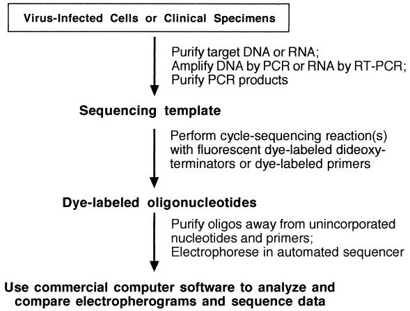
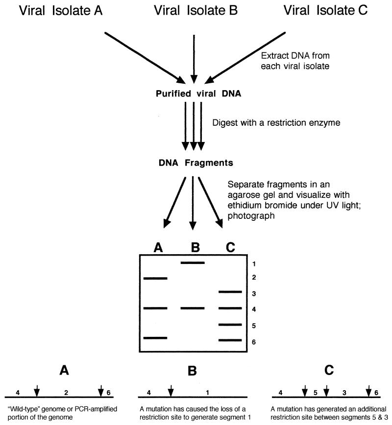
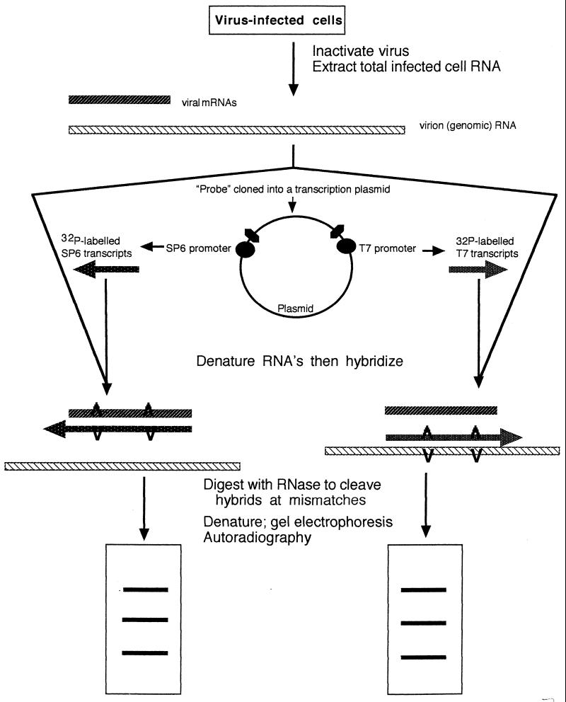
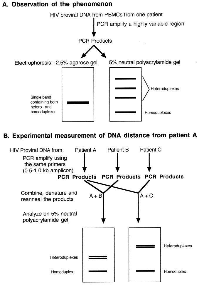
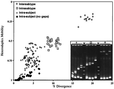
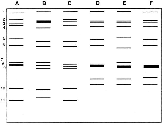
The development over the past two decades of molecular methods for manipulation of RNA and DNA has afforded molecular virologists the ability to study viral genomes in detail that has heretofore not been possible. There are many molecular techniques now available for typing and subtyping of viruses. The available methods include restriction fragment length polymorphism analysis, Southern blot analysis, oligonucleotide fingerprint analysis, reverse hybridization, DNA enzyme immunoassay, RNase protection analysis, single-strand conformation polymorphism analysis, heteroduplex mobility assay, nucleotide sequencing, and genome segment length polymorphism analysis. The methods have certain advantages and disadvantages which should be considered in their application to specific viruses or for specific purposes. These techniques are likely to become more widely used in the future for epidemiologic studies and for investigations into the pathophysiology of virus infections.


Figures

FIG. 1
FIG. 1
Automated cycle sequencing as a rapid and convenient method for sequence analysis of PCR products. The target DNA was extracted and amplified and the PCR products were purified away from unincorporated nucleotides and primers. The sequencing template was “amplified” in another PCR mixture containing the sequencing primers, a mixture of deoxy- and dideoxynucleotides, and modified Taq polymerase. Dideoxynucleotides were coupled to fluorescent dyes, a different color for each of the four bases; these act as terminators of the growing oligonucleotides. Thus, the color of the dye is the key to which dideoxynucleotide is at the 3′ terminus of each oligonucleotide. The large array of oligonucleotides was separated in a polyacrylamide gel which was continuously scanned by a laser during the run, and the colors were detected as they passed the scan point. Each oligonucleotide in the array, as they move through the gel, is only one base shorter than the oligonucleotide above it in the gel. The detection information was fed directly into a computer for analysis and construction of the four-color electropherogram, which was translated into the linear sequence by commercially available software.
FIG. 2
FIG. 2
Hypothetical comparison of three viral isolates by RFLP analysis. In this case, viral genomic DNA was extracted from cells infected with the different isolates and then was digested with a restriction enzyme to produce DNA fragments, which were separated in an agarose gel containing ethidium bromide to allow subsequent visualization and documentation of patterns. Virus isolate A was the hypothetical wild type, with two restriction sites. Isolates B and C differ from A by the loss and gain, respectively, of a restriction site, which resulted in altered banding patterns in the gel. The method can be modified for the use of PCR products instead of genomic DNA or for the use of RT-PCR to allow the use of genomic RNA.
FIG. 3
FIG. 3
RNase protection analysis. RNA from virus-infected cells was extracted and purified. This mixture of mRNA and genomic RNA was mixed with transcripts, labeled in vitro with 32P, from a cloned probe. The cloned probe was transcribed in both directions, from an SP6 promoter at one end and a T7 promoter at the other end. The mixtures were denatured and reannealed to allow the formation of hybrids which would contain “bubbles” at the sites of mismatches. RNase digests all the single-stranded regions and cleaves the double-stranded regions at the bubbles or mismatches. After RNase digestion, the products were subjected to electrophoresis in polyacrylamide gels and bands were detected by autoradiography. Viral isolates with different mutations relative to the probe differ from each other in their banding patterns. Adapted from reference with permission of the publisher.
FIG. 4
FIG. 4
SSCP analysis. RT-PCR products from the 5′ NC regions from various HCV genotypes were electrophoresed in 11.5% polyacrylamide gel and visualized by silver staining. HCV types and subtypes are shown across the top. Reprinted from reference with permission of the publisher.
FIG. 5
FIG. 5
HMA. (A) Illustration of the phenomenon and how HMA was initially observed. PCR products generated from a highly variable region of HIV contain many heteroduplexes which are not resolved in agarose gels but may be resolved and appear as distinct bands in polyacrylamide gels. The heteroduplexes migrate more slowly due to conformational changes which result from the bulged regions and can be converted to homoduplexes as described in the text. PBMCs, peripheral blood mononuclear cells. (B) Illustration of the use of the technique to determine the relative relatedness of virus from two patients to that from patient A. Purified amplicons from the test patients were mixed with amplicons from patient A, denatured, reannealed, and subjected to electrophoresis in a neutral polyacrylamide gel. The migration of the heteroduplex bands was retarded in proportion to their genetic distance from the virus from patient A. See the text for details.
FIG. 6
FIG. 6
Use of HMA to determine HIV relatedness. Heteroduplex mobilities versus DNA distance of sequences of the region of HIV-1 Env from V3 to V5 from the same individuals and from individuals infected with the same (intrasubtype) or different (intersubtype) subtypes of HIV-1. Mobilities were determined by dividing the average distance of migration of the heteroduplex bands by the distance of migration of the homoduplex band. Cloned fragments of the patient proviral DNA were used as starting material. DNA distances (i.e., divergence) were determined by counting mismatches in the best alignment of the sequences, disregarding bases that are unpaired due to insertions or deletions. (Inset) Polyacrylamide (5%) gel stained with ethidium bromide showing representative heteroduplexes from the comparisons discussed above. Courtesy of E. L. Delwart and J. I. Mullins.
FIG. 7
FIG. 7
Electropherotyping of rotaviruses. Stylized drawing of 10% polyacrylamide gel analysis of representative RNAs (A through F) extracted from stool specimens from children. The gels were stained with ethidium bromide to visualize the RNA bands (genome segments) numbered 1 to 11 at the left. Samples A through C are the long electropherotype and samples D through F are the short electropherotype. Adapted from reference .

References

    1. Adams S G, Dohner D E, Gelb L D. Restriction fragment differences between the genomes of the Oka varicella vaccine virus and American wild-type varicella-zoster virus. J Med Virol. 1989;29:38–45. - PubMed
    1. Agostini H T, Ryschkewitsch C F, Mory R, Singer E J, Stoner G L. JC virus (JCV) genotypes in brain tissue from patients with progressive multifocal leukoencephalopathy (PML) and in urine from controls without PML: increased frequency of JCV type 2 in PML. J Infect Dis. 1997;176:1–8. - PubMed
    1. Ando T, Jin Q, Gentsch J R, Monroe S S, Noel J S, Dowell S F, Cicirello H G, Kohn M A, Glass R I. Epidemiologic applications of novel molecular methods to detect and differentiate small round structured viruses (Norwalk-like viruses) J Med Virol. 1995;47:145–152. - PubMed
    1. Arens M, Dilworth V. Remarkably homogeneous population of adenovirus type 3 and 7 genome types. J Clin Microbiol. 1988;26:1604–1608. - PMC - PubMed
    1. Arens M, Swierkosz E M. Detection of rotavirus by hybridization with a nonradioactive synthetic DNA probe and comparison with commercial enzyme immunoassays and silver-stained polyacrylamide gels. J Clin Microbiol. 1989;27:1277–1279. - PMC - PubMed

MeSH terms

LinkOut - more resources