

Cloning and sequence analysis of two Pseudomonas flavoprotein xenobiotic reductases
- PMID: 10515912
- PMCID: PMC103757
- DOI: 10.1128/JB.181.20.6254-6263.1999
Cloning and sequence analysis of two Pseudomonas flavoprotein xenobiotic reductases
Abstract
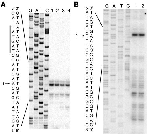
The genes encoding flavin mononucleotide-containing oxidoreductases, designated xenobiotic reductases, from Pseudomonas putida II-B and P. fluorescens I-C that removed nitrite from nitroglycerin (NG) by cleavage of the nitroester bond were cloned, sequenced, and characterized. The P. putida gene, xenA, encodes a 39,702-Da monomeric, NAD(P)H-dependent flavoprotein that removes either the terminal or central nitro groups from NG and that reduces 2-cyclohexen-1-one but did not readily reduce 2,4,6-trinitrotoluene (TNT). The P. fluorescens gene, xenB, encodes a 37,441-Da monomeric, NAD(P)H-dependent flavoprotein that exhibits fivefold regioselectivity for removal of the central nitro group from NG and that transforms TNT but did not readily react with 2-cyclohexen-1-one. Heterologous expression of xenA and xenB was demonstrated in Escherichia coli DH5alpha. The transcription initiation sites of both xenA and xenB were identified by primer extension analysis. BLAST analyses conducted with the P. putida xenA and the P. fluorescens xenB sequences demonstrated that these genes are similar to several other bacterial genes that encode broad-specificity flavoprotein reductases. The prokaryotic flavoprotein reductases described herein likely shared a common ancestor with old yellow enzyme of yeast, a broad-specificity enzyme which may serve a detoxification role in antioxidant defense systems.
Figures


Similar articles
-
Transformation of RDX and other energetic compounds by xenobiotic reductases XenA and XenB.Appl Microbiol Biotechnol. 2009 Sep;84(3):535-44. doi: 10.1007/s00253-009-2024-6. Epub 2009 May 20. Appl Microbiol Biotechnol. 2009. PMID: 19455327 Free PMC article.
-
Cloning, sequencing, and expression of the structural genes for the cytochrome and flavoprotein subunits of p-cresol methylhydroxylase from two strains of Pseudomonas putida.J Bacteriol. 1994 Oct;176(20):6349-61. doi: 10.1128/jb.176.20.6349-6361.1994. J Bacteriol. 1994. PMID: 7929007 Free PMC article.
-
Evaluation of two xenobiotic reductases from Pseudomonas putida for their suitability for magnetic nanoparticle-directed enzyme prodrug therapy as a novel approach to cancer treatment.Microbiologyopen. 2020 Oct;9(10):e1110. doi: 10.1002/mbo3.1110. Epub 2020 Sep 26. Microbiologyopen. 2020. PMID: 32979040 Free PMC article.
-
Regioselectivity of nitroglycerin denitration by flavoprotein nitroester reductases purified from two Pseudomonas species.J Bacteriol. 1997 Nov;179(22):6912-20. doi: 10.1128/jb.179.22.6912-6920.1997. J Bacteriol. 1997. PMID: 9371434 Free PMC article.
-
Generating disulfides in multicellular organisms: emerging roles for a new flavoprotein family.J Biol Chem. 2007 May 11;282(19):13929-33. doi: 10.1074/jbc.R600037200. Epub 2007 Mar 12. J Biol Chem. 2007. PMID: 17353193 Review. No abstract available.
Cited by
-
Genome sequence of the enterobacterial phytopathogen Erwinia carotovora subsp. atroseptica and characterization of virulence factors.Proc Natl Acad Sci U S A. 2004 Jul 27;101(30):11105-10. doi: 10.1073/pnas.0402424101. Epub 2004 Jul 19. Proc Natl Acad Sci U S A. 2004. PMID: 15263089 Free PMC article.
-
Mechanistic Insights into the Ene-Reductase-Catalyzed Promiscuous Reduction of Oximes to Amines.ACS Catal. 2023 Feb 6;13(4):2610-2618. doi: 10.1021/acscatal.2c06137. eCollection 2023 Feb 17. ACS Catal. 2023. PMID: 36846821 Free PMC article.
-
Biotransformation of explosives by the old yellow enzyme family of flavoproteins.Appl Environ Microbiol. 2004 Jun;70(6):3566-74. doi: 10.1128/AEM.70.6.3566-3574.2004. Appl Environ Microbiol. 2004. PMID: 15184158 Free PMC article.
-
Transcriptome profiling defines a novel regulon modulated by the LysR-type transcriptional regulator MexT in Pseudomonas aeruginosa.Nucleic Acids Res. 2009 Dec;37(22):7546-59. doi: 10.1093/nar/gkp828. Nucleic Acids Res. 2009. PMID: 19846594 Free PMC article.
-
Transcriptomics Indicates Active and Passive Metronidazole Resistance Mechanisms in Three Seminal Giardia Lines.Front Microbiol. 2017 Mar 17;8:398. doi: 10.3389/fmicb.2017.00398. eCollection 2017. Front Microbiol. 2017. PMID: 28367140 Free PMC article.
References
-
- Åkeson Å, Ehrenberg A, Theorell H. Old yellow enzyme. In: Boyer P D, Lardy H, Myrbäck K, editors. The enzymes. New York, N.Y: Academic Press; 1963. pp. 339–416.
-
- Baca M, Borgstahl G E, Boissinot M, Burke P M, Williams D R, Slater K A, Getzoff E D. Complete chemical structure of photoactive yellow protein: novel thioester-linked 4-hydroxycinnamyl chromophore and photocycle. Biochemistry. 1994;33:14369–14377. - PubMed
Publication types
MeSH terms
Substances
Associated data
- Actions
- Actions
LinkOut - more resources
Full Text Sources
Other Literature Sources
Molecular Biology Databases
Research Materials
