

Mutation analysis of the 5' untranslated region of the cold shock cspA mRNA of Escherichia coli
- PMID: 10515916
- PMCID: PMC103761
- DOI: 10.1128/JB.181.20.6284-6291.1999
Mutation analysis of the 5' untranslated region of the cold shock cspA mRNA of Escherichia coli
Abstract
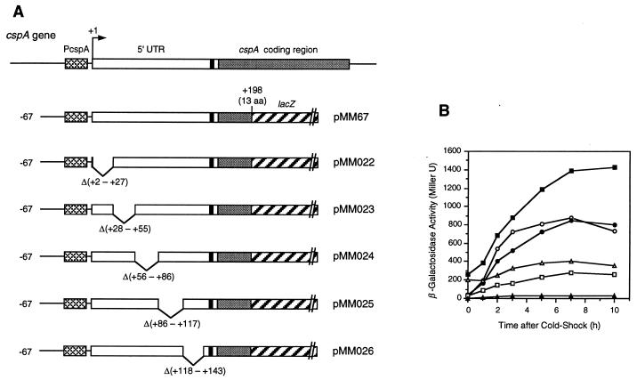
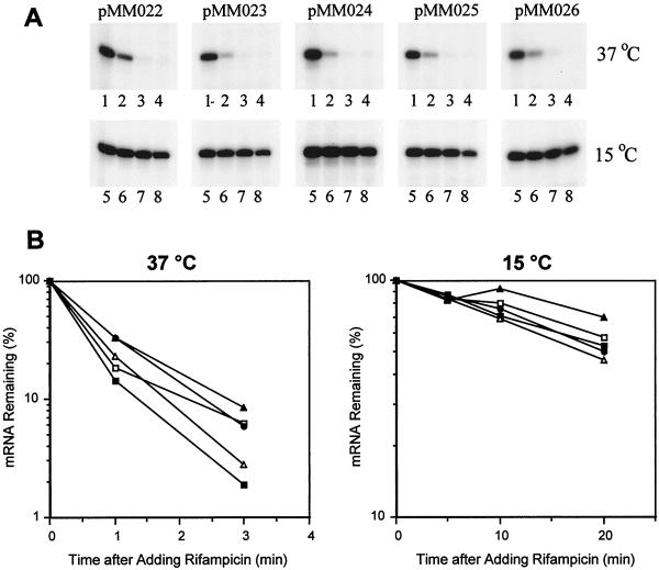
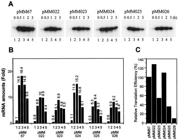
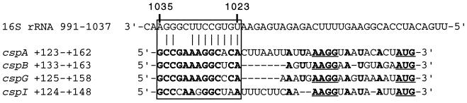
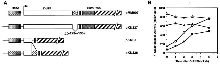
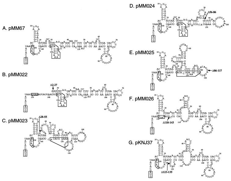
The mRNA for CspA, a major cold shock protein in Escherichia coli, contains an unusually long (159 bases) 5' untranslated region (5'-UTR), and its stability has been shown to play a major role in cold shock induction of CspA. The 5'-UTR of the cspA mRNA has a negative effect on its expression at 37 degrees C but has a positive effect upon cold shock. In this report, a series of cspA-lacZ fusions having a 26- to 32-base deletion in the 5'-UTR were constructed to examine the roles of specific regions within the 5'-UTR in cspA expression. It was found that none of the deletion mutations had significant effects on the stability of mRNA at both 37 and 15 degrees C. However, two mutations (Delta56-86 and Delta86-117) caused a substantial increase of beta-galactosidase activity at 37 degrees C, indicating that the deleted regions contain a negative cis element(s) for translation. A mutation (Delta2-27) deleting the highly conserved cold box sequence had little effect on cold shock induction of beta-galactosidase. Interestingly, three mutations (Delta28-55, Delta86-117, and Delta118-143) caused poor cold shock induction of beta-galactosidase. In particular, the Delta118-143 mutation reduced the translation efficiency of the cspA mRNA to less than 10% of that of the wild-type construct. The deleted region contains a 13-base sequence named upstream box (bases 123 to 135), which is highly conserved in cspA, cspB, cspG, and cspI, and is located 11 bases upstream of the Shine-Dalgarno (SD) sequence. The upstream box might be another cis element involved in translation efficiency of the cspA mRNA in addition to the SD sequence and the downstream box sequence. The relationship between the mRNA secondary structure and translation efficiency is discussed.
Figures
Similar articles
-
CspI, the ninth member of the CspA family of Escherichia coli, is induced upon cold shock.J Bacteriol. 1999 Mar;181(5):1603-9. doi: 10.1128/JB.181.5.1603-1609.1999. J Bacteriol. 1999. PMID: 10049393 Free PMC article.
-
Role of the cold-box region in the 5' untranslated region of the cspA mRNA in its transient expression at low temperature in Escherichia coli.J Bacteriol. 1998 Jan;180(1):90-5. doi: 10.1128/JB.180.1.90-95.1998. J Bacteriol. 1998. PMID: 9422597 Free PMC article.
-
The role of the 5'-end untranslated region of the mRNA for CspA, the major cold-shock protein of Escherichia coli, in cold-shock adaptation.J Bacteriol. 1996 Aug;178(16):4919-25. doi: 10.1128/jb.178.16.4919-4925.1996. J Bacteriol. 1996. PMID: 8759856 Free PMC article.
-
The CspA family in Escherichia coli: multiple gene duplication for stress adaptation.Mol Microbiol. 1998 Jan;27(2):247-55. doi: 10.1046/j.1365-2958.1998.00683.x. Mol Microbiol. 1998. PMID: 9484881 Review.
-
The cold-shock response--a hot topic.Mol Microbiol. 1994 Mar;11(5):811-8. doi: 10.1111/j.1365-2958.1994.tb00359.x. Mol Microbiol. 1994. PMID: 8022259 Review.
Cited by
-
Characterization of the groEL and groES loci in Bifidobacterium breve UCC 2003: genetic, transcriptional, and phylogenetic analyses.Appl Environ Microbiol. 2004 Oct;70(10):6197-209. doi: 10.1128/AEM.70.10.6197-6209.2004. Appl Environ Microbiol. 2004. PMID: 15466567 Free PMC article.
-
A tight cold-inducible switch built by coupling thermosensitive transcriptional and proteolytic regulatory parts.Nucleic Acids Res. 2019 Dec 2;47(21):e137. doi: 10.1093/nar/gkz785. Nucleic Acids Res. 2019. PMID: 31750522 Free PMC article.
-
Microbial thermosensors.Cell Mol Life Sci. 2009 Aug;66(16):2661-76. doi: 10.1007/s00018-009-0041-3. Epub 2009 May 12. Cell Mol Life Sci. 2009. PMID: 19554260 Free PMC article. Review.
-
Selective mRNA degradation by polynucleotide phosphorylase in cold shock adaptation in Escherichia coli.J Bacteriol. 2001 May;183(9):2808-16. doi: 10.1128/JB.183.9.2808-2816.2001. J Bacteriol. 2001. PMID: 11292800 Free PMC article.
-
Enhanced synthesis of internalin A in aro mutants of Listeria monocytogenes indicates posttranscriptional control of the inlAB mRNA.J Bacteriol. 2005 Apr;187(8):2836-45. doi: 10.1128/JB.187.8.2836-2845.2005. J Bacteriol. 2005. PMID: 15805530 Free PMC article.
References
-
- Bae W, Phadtare S, Severinov K, Inouye M. Characterization of Escherichia coli cspE, whose product negatively regulates transcription of cspA, the gene for the major cold shock protein. Mol Microbiol. 1999;31:1429–1441. - PubMed
-
- Brandi A, Pietroni P, Gualerzi C O, Pon C L. Post-transcriptional regulation of CspA expression in Escherichia coli. Mol Microbiol. 1996;19:231–240. - PubMed
Publication types
MeSH terms
Substances
Grants and funding
LinkOut - more resources
Full Text Sources
Other Literature Sources
