

MinDE-dependent pole-to-pole oscillation of division inhibitor MinC in Escherichia coli
- PMID: 10515933
- PMCID: PMC103778
- DOI: 10.1128/JB.181.20.6419-6424.1999
MinDE-dependent pole-to-pole oscillation of division inhibitor MinC in Escherichia coli
Abstract
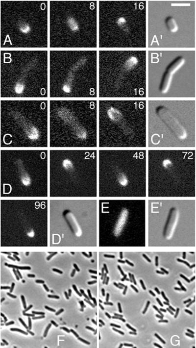
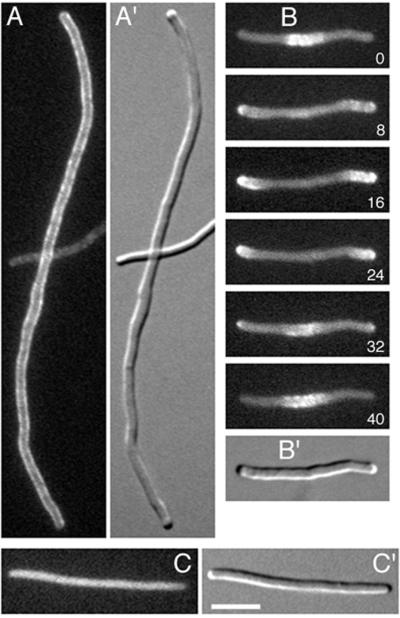
By inhibiting FtsZ ring formation near the cell ends, the MinC protein plays a critical role in proper positioning of the division apparatus in Escherichia coli. MinC activity requires that of MinD, and the MinE peptide provides topological specificity by suppressing MinC-MinD-mediated division inhibition specifically at the middle of the cell. We recently presented evidence that MinE not only accumulates in an FtsZ-independent ring structure at the cell's middle but also imposes a unique dynamic localization pattern upon MinD in which the latter accumulates alternately in either one of the cell halves in what appears to be a rapidly oscillating membrane association-dissociation cycle. Here we show that functional green fluorescent protein-MinC displays a very similar oscillatory behavior which is dependent on both MinD and MinE and independent of FtsZ. The results support a model in which MinD recruits MinC to its site of action and in which FtsZ ring assembly at each of the cell ends is blocked in an intermittent and alternate fashion.
Figures


 , MinD;
  , the MinE ring. PDSs are represented by either a minus (blocked by MinC-MinD) or a plus (not blocked, available for FtsZ ring assembly) sign. (A) In the absence of MinD and MinE, MinC localizes nonspecifically to the cytoplasm and has no effect on septal or FtsZ ring formation. (B) In the presence of MinD, and absence of MinE, MinC associates with MinD along the entire membrane, preventing FtsZ ring formation at all PDSs. (C) In WT cells, MinC co-oscillates with MinD from one side of the MinE ring to the other, actively interfering with FtsZ ring assembly at each cell end in a sequential and rapidly repeating fashion. (D) In cells lacking FtsZ rings, multiple MinE rings define three or more cell segments. As in WT cells, MinC co-oscillates with MinD between the segments flanking each MinE ring. Note that although the figure suggests the presence of a limited number of regularly spaced PDSs in each cell, the proposed mechanism of MinCDE action does not depend on the exact number or nature of the PDSs and is equally tenable whether potential sites for FtsZ ring assembly are (co)determined by positioning of the nucleoids (41) or any other mechanism.
References
Publication types
MeSH terms
Substances
Grants and funding
LinkOut - more resources
Full Text Sources
Other Literature Sources
Molecular Biology Databases
