. 1999 Nov;73(11):8950-7.
doi: 10.1128/JVI.73.11.8950-8957.1999.

Herpes simplex virus inhibits apoptosis through the action of two genes, Us5 and Us3

Affiliations

Herpes simplex virus inhibits apoptosis through the action of two genes, Us5 and Us3

K R Jerome et al. J Virol. 1999 Nov.

Abstract

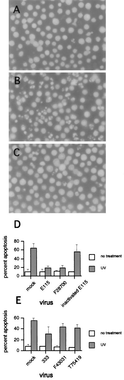
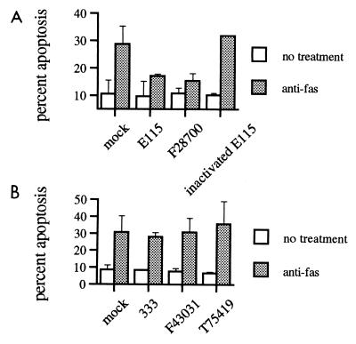
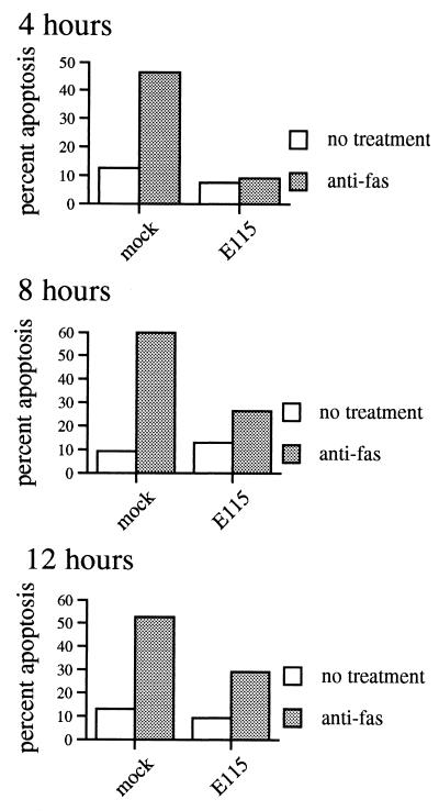
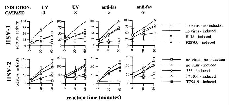
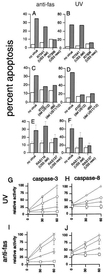
Apoptosis of virus-infected cells occurs either as a direct response to viral infection or upon recognition of infection by the host immune response. Apoptosis reduces production of new virus from these cells, and therefore viruses have evolved inhibitory mechanisms. We previously showed that laboratory strains of herpes simplex virus type 1 (HSV-1) protect infected cells from apoptosis induced by cytotoxic T lymphocytes or ethanol. We have now evaluated the ability of HSV-1 and HSV-2 laboratory and clinical isolates to inhibit apoptosis induced by anti-Fas antibody or UV irradiation and explored the genetic basis for this inhibition. HSV-1 isolates inhibited apoptosis induced by UV or anti-Fas antibody. In contrast, HSV-2 clinical isolates failed to inhibit apoptosis induced by either stimulus, although the HSV-2 laboratory strain 333 had a partial inhibitory effect on UV-induced apoptosis. Inhibition of apoptosis by HSV was accompanied by marked reduction of caspase-3 and caspase-8 activity. Deletion of the HSV-1 Us3 gene markedly reduced inhibition of UV-induced apoptosis and partially abrogated inhibition of Fas-mediated apoptosis. Conversely, deletion of the HSV-1 Us5 gene markedly reduced protection from Fas-mediated apoptosis and partially abrogated protection from UV. The Us11 and Us12 genes were not necessary for protection from apoptosis induced by either stimulus. The differences between HSV-1 and HSV-2 in the ability to inhibit apoptosis may be factors in the immunobiology of HSV infections.


Figures

FIG. 1
FIG. 1
Clinical and laboratory strains of HSV-1 and HSV-2 inhibit UV-induced apoptosis. Jurkat cells were mock or HSV infected for 5 h prior to induction of apoptosis with UV radiation. The morphological changes of apoptosis were assessed by a blinded observer using fluorescence microscopy. (A) Uninfected cells without UV irradiation; (B) uninfected cells after UV irradiation; (C) HSV-1-infected cells (strain H61146) after treatment with UV; (D and E) percent apoptosis after infection with a clinical (F28700, F43031, and T75419) or laboratory (E115 and 333) strains of HSV-1 (D) and HSV-2 (E). Shown is mean + SEM of two to five independent experiments. In some instances, error bars are too small to be seen.
FIG. 2
FIG. 2
Strains of HSV-1, but not HSV-2, inhibit Fas-mediated apoptosis. Percent apoptosis by anti-Fas antibody APO-1-3 after infection with clinical (F28700, F43031, and T75419) or laboratory (E115 and 333) strains of HSV-1 (A) or HSV-2 (B), as determined by a blinded observer using fluorescence microscopy. Shown is mean + SEM of two to five independent experiments. In some instances, error bars are too small to be seen.
FIG. 3
FIG. 3
Time course of HSV inhibition of Fas-mediated apoptosis. Jurkat cells were mock or HSV-1 infected, incubated for 5 h, and treated with 1,000 ng of anti-Fas antibody CH11 per ml. Apoptosis was evaluated at the indicated times after anti-Fas treatment by a blinded observer using fluorescence microscopy. Cells could not be evaluated at 24 h due to extensive viral cytopathic effect. Shown is mean of two independent experiments.
FIG. 4
FIG. 4
Inhibition of caspase activity after UV or anti-Fas treatment by HSV-1 and HSV-2. Jurkat cells were mock or HSV infected for 5 h prior to induction of apoptosis with UV radiation or anti-Fas antibody. Caspase-3 (-8) or caspase-8 (-8) activity was measured in cell lysates made 4 h later. Virus infection alone with HSV-1 or HSV-2 did not lead to caspase-3 or caspase-8 activation (not shown). Shown is mean ± SEM of from two to five independent experiments. In some instances, error bars are too small to be seen.
FIG. 5
FIG. 5
Inhibition of Fas-mediated or UV-induced apoptosis by deletion mutants of HSV-1. Jurkat cells were infected with the indicated mutant virus at 10 PFU/cell for 5 h, and apoptosis was induced by anti-Fas antibody (A, C, and E) or UV irradiation (B, D, and F). Open bars, untreated cells; shaded bars, treated cells. Percent apoptosis was determined by a blinded observer using fluorescence microscopy. Shown is the mean + SEM of two to four independent experiments. (G to J) Inhibition of caspase activity by HSV-1 deletion mutant RAS116 (○) and rescue mutant RAS137 (▵). Jurkat cells were mock infected (◊) or HSV infected for 5 h prior to induction of apoptosis with UV radiation or anti-Fas antibody. Caspase-3 or caspase-8 activity was measured in cell lysates made 4 h. Shown is mean ± SEM of two to five independent experiments. In some instances, error bars are too small to be seen. □, uninduced mock-infected cells.
FIG. 6
FIG. 6
One-step growth curve of HSV in Jurkat cells. Jurkat cells were infected with HSV-1 (F28700) or HSV-2 (F43031) at 5 PFU/cell. Infected cultures were either left unirradiated or UV-irradiated at 2 or 5 h postinfection. Viral titers at the indicated time points were determined by a standard plaque assay.

References

    1. Aragane Y, Kulms D, Metze D, Wilkes G, Poppelmann B, Luger T A, Schwartz T. Ultraviolet light induces apoptosis via direct activation of CD95 (Fas/APO-1) independently of its ligand CD95L. J Cell Biol. 1998;140:171–182. - PMC - PubMed
    1. Ashkenazi A, Dixit V M. Death receptors: signaling and modulation. Science. 1998;281:1305–1308. - PubMed
    1. Aubert M, Blaho J A. The herpes simplex type 1 regulatory protein ICP27 is required for the prevention of apoptosis in infected human cells. J Virol. 1999;73:2803–2813. - PMC - PubMed
    1. Balan P, Davis-Poynter N, Bell S, Atkinson H, Browne H, Minson T. An analysis of the in vitro and in vivo phenotypes of mutants of herpes simplex virus type 1 lacking glycoproteins gG, gE, gI or the putative gJ. J Gen Virol. 1994;75:1245–1258. - PubMed
    1. Barry M, McFadden G. Apoptosis regulators from DNA viruses. Curr Opin Immunol. 1998;10:422–430. - PubMed

Publication types

MeSH terms