. 1999 Nov;73(11):9137-44.
doi: 10.1128/JVI.73.11.9137-9144.1999.

Replication-defective bovine adenovirus type 3 as an expression vector

Affiliations

Replication-defective bovine adenovirus type 3 as an expression vector

P S Reddy et al. J Virol. 1999 Nov.

Abstract

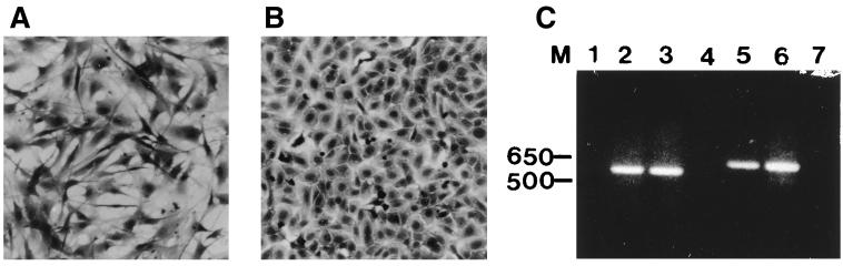
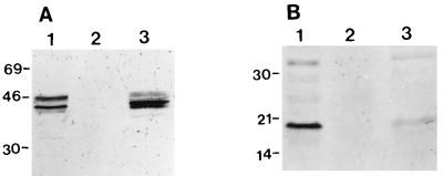
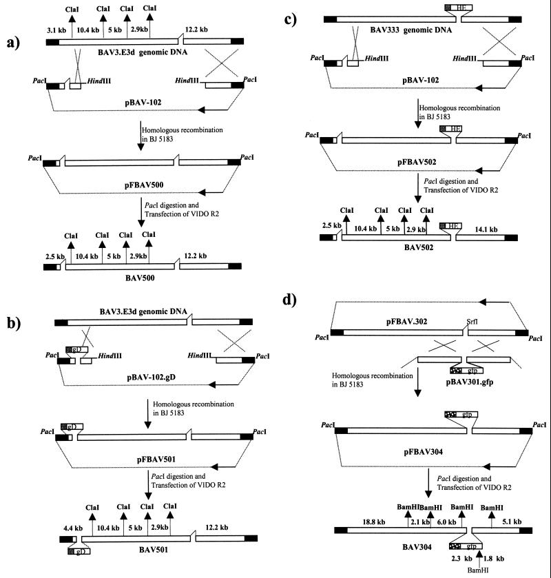
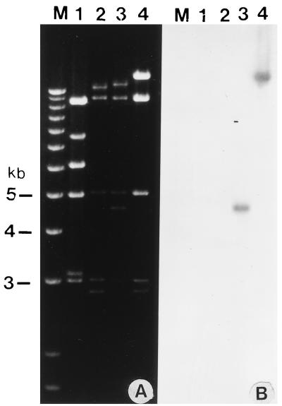
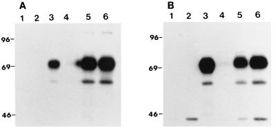
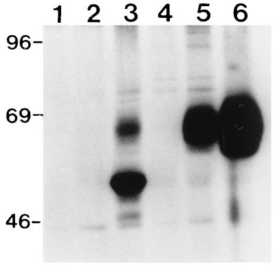
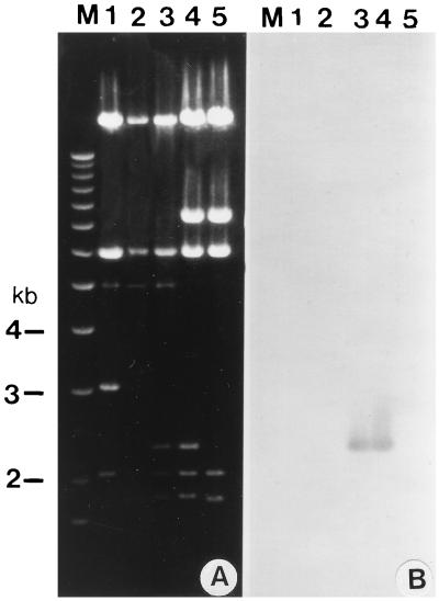
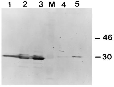
Although recombinant human adenovirus (HAV)-based vectors offer several advantages for somatic gene therapy and vaccination over other viral vectors, it would be desirable to develop alternative vectors with prolonged expression and decreased toxicity. Toward this objective, a replication-defective bovine adenovirus type 3 (BAV-3) was developed as an expression vector. Bovine cell lines designated VIDO R2 (HAV-5 E1A/B-transformed fetal bovine retina cell [FBRC] line) and 6.93.9 (Madin-Darby bovine kidney [MDBK] cell line expressing E1 proteins) were developed and found to complement the E1A deletion in BAV-3. Replication-defective BAV-3 with a 1.7-kb deletion removing most of the E1A and E3 regions was constructed. This virus could be grown in VIDO R2 or 6.93.9 cells but not in FBRC or MDBK cells. The results demonstrated that the E1 region of HAV-5 has the capacity to transform bovine retina cells and that the E1A region of HAV-5 can complement that of BAV-3. A replication-defective BAV-3 vector expressing bovine herpesvirus type 1 glycoprotein D from the E1A region was made. A similar replication-defective vector expressing the hemagglutinin-esterase gene of bovine coronavirus from the E3 region was isolated. Although these viruses grew less efficiently than the replication-competent recombinant BAV-3 (E3 deleted), they are suitable for detailed studies with animals to evaluate the safety, duration of foreign gene expression, and ability to induce immune responses. In addition, a replication-competent recombinant BAV-3 expressing green fluorescent protein was constructed and used to evaluate the host range of BAV-3 under cell culture conditions. The development of bovine E1A-complementing cell lines and the generation of replication-defective BAV-3 vectors is a major technical advancement for defining the use of BAV-3 as vector for vaccination against diseases of cattle and somatic gene therapy in humans.


Figures

FIG. 1
FIG. 1
VIDO R2 cell line. Morphological features of untransformed FBRCs (A) and transformed VIDO R2 cells (B). Cells were stained with crystal violet and photographed at a magnification of ×100. (C) Analysis of ethidium bromide-stained RT-PCR products. Products of RT-PCR using DNase-treated RNA (lanes 1, 2, 4, 5, and 7) or of PCR using DNA (lanes 3 and 6) as a template were synthesized by using primer pairs for E1A (sense, 5′-ATGAGACATATTATCTGCCA-3′; antisense, 5′-CTTACTGTAGACAAACATGC) (lanes 1 to 3) and 19-kDa E1B (sense, 5′-ATGTTTAACTTGCATGGCGT-3′; antisense, 5′-ATTCCCGAGGGTCCAGGCCG-3′) (lanes 4 to 6). Lanes 1 (E1A primers) and 4 (19-kDa E1B primers) shows RT-PCR of RNA without reverse transcriptase; lane 7 shows RT-PCR using RNA from FBRCs. Sizes of marker (M) DNA are shown in base pairs.
FIG. 2
FIG. 2
Western blot analysis of E1 proteins. Proteins from 293 cells (lane 1), FBRCs (lane 2), and VIDO R2 cells (lane 3) were separated by SDS-PAGE (10% gel) under reducing conditions and transferred to nitrocellulose. The separated proteins were probed in Western blots by MAbs against HAV-5 E1A (A) and 19-kDa E1B (B). Sizes are indicated in kilodaltons.
FIG. 3
FIG. 3
Strategy used for the generation of recombinant BAV-3. Different plasmids were constructed from different genomic clones as described in the text. Origins of DNA sequences: plasmid DNA, thin line; BAV-3 genomic DNA, hollow box; inverted terminal repeats, filled box. The plasmid maps are not drawn to scale.
FIG. 4
FIG. 4
Restriction enzyme analysis of the recombinant BAV-3 genome. (A) The DNAs were extracted from VIDO R2 cells infected with BAV-3 (lane 1), BAV500 (lane 2), BAV501 (lane 3), or BAV502 (lane 4) as described previously (17) and digested with ClaI. (B) The fragments shown in panel A were transferred to Nytran membranes and probed with α-32P-labeled gD and HE probes. Lane M, 1 Kb Plus DNA Ladder (Gibco/BRL) used for sizing the viral DNA fragments.
FIG. 5
FIG. 5
Production of recombinant BAV-3 by VIDO R2 and FBRC cells. Near-confluent monolayers were infected with less than 1 MOI of wild-type or recombinant BAV-3. After a week, the cells were freeze-thawed and the virus was titrated on VIDO R2 cells as described in the text.
FIG. 6
FIG. 6
Kinetics of gD expression by recombinant BAV-3. Proteins from lysates of [35S]methionine-labeled mock-infected (lane 1), BAV-3-infected (lane 2), BHV-1-infected (lane 3), or BAV501-infected cells harvested at 12 (lane 4), 24 (lane 5), and 36 (lane 6) h postinfection were immunoprecipitated with anti-gD MAbs (18) and separated by SDS-PAGE 10% gel under reducing conditions. (A) VIDO R2 cells; (B) MDBK cells. Positions of size markers (in kilodaltons) are shown to the left of each panel.
FIG. 7
FIG. 7
Kinetics of HE expression by recombinant BAV-3 in VIDO R2 cells. Proteins from lysates of [35S]methionine-labeled mock-infected (lane 1), BAV-3-infected (lane 2), BCV-infected (lane 3), or BAV502-infected cells harvested at 12 (lane 4), 24 (lane 5), and 36 (lane 6) h postinfection were immunoprecipitated with anti-BCV polyclonal antibodies (9, 10) and separated by SDS-PAGE (10% gel) under reducing conditions. Positions of size markers (in kilodaltons) are shown on the left.
FIG. 8
FIG. 8
Analysis of recombinant BAV304 genome. (A) The DNAs were extracted from BAV-3 (lane 1), BAV3.E3d (lane 2), BAV304 (lane 3), pFBAV304 (lane 4), and pFBAV302 (lane 5) digested with BamHI and analyzed by ethidium bromide staining of an agarose gel. (B) The fragments shown in panel A were transferred to a Nytran membrane and probed with a α-32P-labeled GFP gene. Lane M, 1 Kb Plus DNA Ladder (Gibco/BRL) used for sizing the DNA fragments.
FIG. 9
FIG. 9
Western blot analysis of GFP expression. Proteins from mock (lane 4)-infected, BAV-3-infected (lane 5), or BAV304-infected MDBK cells harvested at 12 (lane 1), 24 (lane 2), and 36 (lane 3) h postinfection were separated by SDS-PAGE (10% gel) under reducing conditions and transferred to nitrocellulose. The separated proteins were probed in Western blots by GFP-specific polyclonal antiserum. Molecular masses (lane M) are indicated in kilodaltons.

References

    1. Baca-Estrada M E, Liang X, Babiuk L A, Yoo D. Induction of mucosal immunity in cotton rats to hamagglutinin-esterase glycoprotein of bovine coronavirus by recombinant adenovirus. Immunology. 1995;86:134–140. - PMC - PubMed
    1. Ball A O, Williams M E, Spindler K R. Identification of mouse adenovirus type 1 early region 1: DNA sequence and a conserved transactivating function. J Virol. 1988;62:3947–3957. - PMC - PubMed
    1. Bartha A. Proposal for subgrouping of bovine adenoviruses. Acta Vet. 1969;19:319–321. - PubMed
    1. Bett A J, Haddara W, Prevec L, Graham F L. An efficient and flexible system for construction of adenovirus vectors with insertions or deletions in early regions 1 and 3. Proc Natl Acad Sci USA. 1994;91:8802–8806. - PMC - PubMed
    1. Chartier C, Degryse E, Gantzer M, Dieterle A, Pavirani A, Mehtali M. Efficient generation of recombinant adenovirus vectors by homologous recombination in Escherichia coli. J Virol. 1996;70:4805–4810. - PMC - PubMed

Publication types

MeSH terms