. 1999 Nov;73(11):9222-31.
doi: 10.1128/JVI.73.11.9222-9231.1999.

Picornavirus internal ribosome entry site elements target RNA cleavage events induced by the herpes simplex virus virion host shutoff protein

Affiliations

Picornavirus internal ribosome entry site elements target RNA cleavage events induced by the herpes simplex virus virion host shutoff protein

M M Elgadi et al. J Virol. 1999 Nov.

Abstract

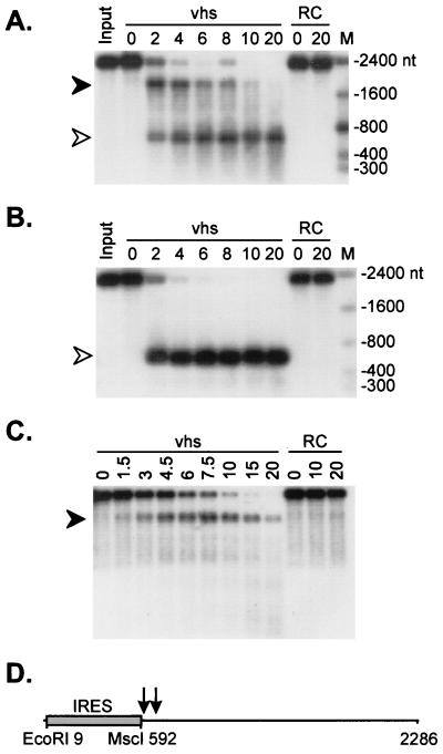
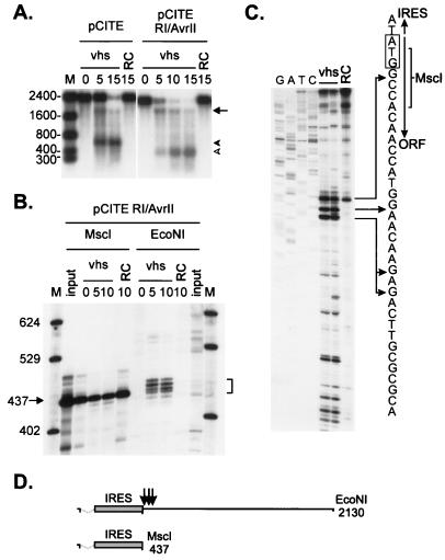
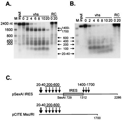
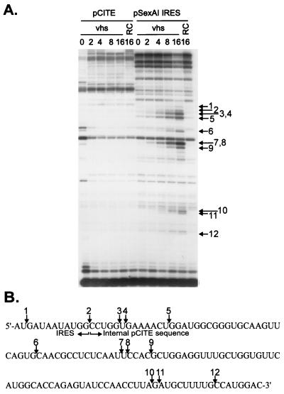
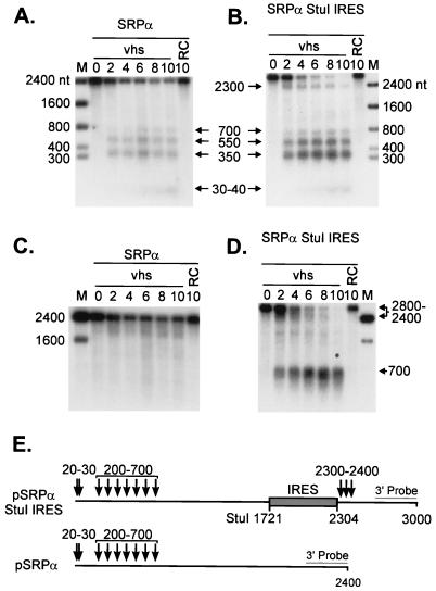
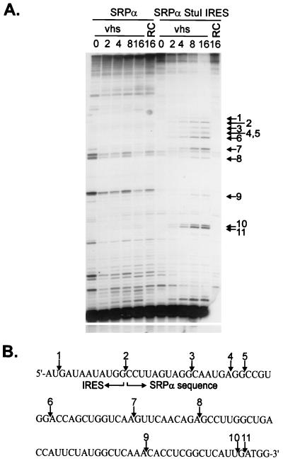
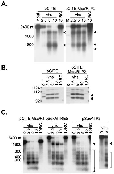
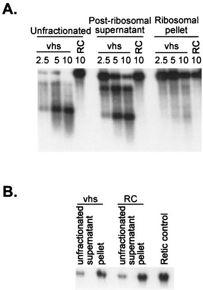
The herpes simplex virus (HSV) virion host shutoff (vhs) protein (UL41 gene product) is a component of the HSV virion tegument that triggers shutoff of host protein synthesis and accelerated mRNA degradation during the early stages of HSV infection. vhs displays weak amino acid sequence similarity to the fen-1 family of nucleases and suffices to induce accelerated RNA turnover through endoribonucleolytic cleavage events when it is expressed as the only HSV protein in a rabbit reticulocyte in vitro translation system. Although vhs selectively targets mRNAs in vivo, the basis for this selectivity remains obscure, since in vitro activity is not influenced by the presence of a 5' cap or 3' poly(A) tail. Here we show that vhs activity is greatly altered by placing an internal ribosome entry site (IRES) from encephalomyocarditis virus or poliovirus in the RNA substrate. Transcripts bearing the IRES were preferentially cleaved by the vhs-dependent endoribonuclease at multiple sites clustered in a narrow zone located immediately downstream of the element in a reaction that did not require ribosomes. Targeting was observed when the IRES was located at the 5' end or placed at internal sites in the substrate, indicating that it is independent of position or sequence context. These data indicate that the vhs-dependent nuclease can be selectively targeted by specific cis-acting elements in the RNA substrate, possibly through secondary structure or a component of the translational machinery.


Figures

FIG. 1
FIG. 1
vhs induces preferential endonucleolytic cleavage in the vicinity of the 3′ boundary of the EMCV IRES. A 2.3-kb runoff transcript of pCITE-1 was added to RRLs containing pretranslated vhs (lanes marked vhs) and to control RRL (lanes marked RC), and samples were removed at the indicated times (in minutes). RNA was extracted from each sample, resolved on a 1% agarose–6% formaldehyde gel, and transferred to a Nytran Plus membrane. (A) Capped, internally labeled RNA. The closed and open arrowheads indicate the ca. 1,800- and 600-nt fragments, respectively. (B) 5′-Cap-labeled RNA. (C) Capped, unlabeled RNA. The radioactive signals in panels A and B were detected by autoradiography. The RNA products in panel C were detected by hybridization to a 5′-labeled oligonucleotide probe complementary to residues 729 to 746 of the pCITE transcript, followed by autoradiography. The numbers to the right of panels A and B indicate the sizes of RNA markers (lanes marked M) in nucleotides. (D) Diagram of the pCITE transcript indicating the approximate position(s) of the cleavage site(s) (arrows).
FIG. 2
FIG. 2
The pCITE transcript is cleaved immediately downstream of the IRES. (A) Uncapped, internally labeled transcripts of pCITE and pCITE RI/AvrII (lacking the 5′-most 159 nt of the IRES) were added to RRLs containing vhs, and the reaction products were analyzed as for Fig. 1A. The arrow and arrowheads indicate the 3′ and 5′ degradation products, respectively. The numbers to the left indicate the sizes of RNA markers (lane M) in nucleotides. RC, control RRL. (B) 5′-Cap-labeled runoff transcripts of pCITE RI/AvrII were added to RRL containing vhs, and reaction products extracted at the indicated times (in minutes) were resolved on an 8% polyacrylamide sequencing gel. The template DNA was linearized with MscI or EcoNI prior to transcription, generating runoff transcripts of 437 and 2,127 nt, respectively. The numbers on the left indicate the sizes of DNA markers (lane M) in nucleotides. The bracket indicates the four prominent 5′ products. (C) A capped, unlabeled EcoNI runoff transcript of pCITE was incubated for 10 min in RRL containing pretranslated vhs or in control RRL (RC). The RNA was then extracted and analyzed by primer extension, using a 5′-labeled oligonucleotide complementary to residues 685 to 710 of the transcript. The primer extension products were resolved on an 8% polyacrylamide sequencing gel along with a DNA sequencing ladder generated from pCITE-1 DNA by using the same primer. The diagram accompanying panel C shows the nucleotide sequence at the IRES/open reading frame boundary. The EMCV translational initiation codon is indicated by a rectangle, and arrows indicate the MscI cleavage site and the positions of some of the 5′ ends generated by vhs-induced cleavage. (D) Structures of the pCITE RI/AvrII runoff transcripts. Dashed lines indicate the extent of the IRES deletion, and arrows indicate the approximate locations of the vhs-dependent cleavage sites.
FIG. 3
FIG. 3
The EMCV IRES serves as a movable targeting element for vhs-induced RNA cleavage. 5′-Cap-labeled EcoNI runoff transcripts of pSexAI IRES (A) and parental pCITE Msc/RI (B) were added to RRLs containing vhs (lanes marked vhs) and to control RRL (lanes marked RC), and samples removed at the indicated times (in minutes) were analyzed by agarose-formaldehyde gel electrophoresis as for Fig. 1. Arrows and associated numbers indicate the positions and sizes (in nucleotides) of the vhs-dependent RNA cleavage products. The numbers to the left indicate the sizes of RNA markers (lanes M) in nucleotides. (C) Diagram showing the structures of the two transcripts and the approximate locations of the vhs-induced cleavages. The shaded box represents the EMCV IRES.
FIG. 4
FIG. 4
vhs induces cleavage downstream of an internally located EMCV IRES. (A) Uncapped, unlabeled transcripts of pCITE and pSexAI IRES were added to RRLs containing vhs (lanes marked vhs) and to control RRL (lanes marked RC), and samples were removed at the indicted times (in minutes). Following extraction, the reaction products were analyzed by primer extension, using a 5′-labeled oligonucleotide complementary to residues 1447 to 1471 of the pSexAI IRES RNA (and residues 1438 to 1462 of the pCITE transcript). Primer extension products were analyzed on an 8% polyacrylamide sequencing gel. Numbered arrows indicate primer extension products resulting from vhs-induced cleavage. (B) Diagram showing the nucleotide sequence at the 3′ boundary of the IRES in pSexAI IRES RNA. Numbered arrows correspond to those in panel A and indicate the locations of the novel 5′ ends produced by vhs-induced cleavage.
FIG. 5
FIG. 5
The EMCV IRES targets vhs-induced cleavage of SRPα mRNA. Cap-labeled SRPα (A) and SRPα StuI IRES (B) RNAs were added to RRLs containing vhs (diluted 1:4 in naive RRL) (lanes marked vhs) and to control RRL (lanes marked RC), and RNA extracted from samples removed at the indicated times (in minutes) was analyzed by agarose-formaldehyde gel electrophoresis as for Fig. 1. (C D) The membranes displayed in panels A and B, respectively, were hybridized to a probe corresponding to the 3′-most 400 nt of SRPα RNA (after the radioactive signal from the cap label had been allowed to decay for more than 6 half-lives). Arrows and associated numbers indicate the positions and sizes of the vhs-induced cleavage intermediates. The numbers to the left of panels A and C and to the right of panels B and D indicate the sizes of RNA markers (lanes M) in nucleotides. (E) Diagram showing the structures of both transcripts, with the approximate locations of vhs-induced cleavages indicated by arrows. The shaded rectangle represents the EMCV IRES.
FIG. 6
FIG. 6
The EMCV IRES targets cleavage to 3′-flanking SRPα sequences. (A) Uncapped, unlabeled SRPα and SRPα StuI IRES RNAs were combined with RRLs containing vhs (lanes marked vhs) and with control RRL (lanes marked RC), and samples were withdrawn at the indicated times (in minutes). RNAs were then extracted and analyzed by primer extension, using a 5′-labeled oligonucleotide that anneals to residues 1841 to 1865 of SRPα RNA (and residues 2424 to 2448 of the SRPα StuI IRES transcript). Numbered arrows indicate the positions of novel 5′ ends resulting from vhs-induced RNA cleavage. (B) Diagram showing the nucleotide sequence at the 3′ boundary of the IRES in SRPα StuI IRES RNA. Numbered arrows correspond to those in panel A and indicate the locations of the vhs-induced cleavage sites.
FIG. 7
FIG. 7
The poliovirus IRES serves as a movable targeting element for vhs-induced RNA cleavage. (A) Uncapped, internally labeled pCITE and pCITE Msc/RI P2 RNAs were reacted with RRLs containing vhs and with control RRL (RC), and samples removed at the indicated times (in minutes) were analyzed by agarose-formaldehyde gel electrophoresis as for Fig. 1. pCITE Msc/RI P2 is a derivative of pCITE in which the EMCV IRES has been replaced by the IRES of poliovirus type 2. (B) Uncapped, unlabeled pCITE and pCITE Msc/RI P2 RNAs were added to RRLs containing vhs and to control RRL. RNAs extracted from samples removed at the indicated times (in minutes) were analyzed by primer extension, using a 5′-labeled oligonucleotide that anneals to residues 685 to 710 of pCITE RNA (and residues ca. 685 to 710 of pCITE Msc/RI P2 RNA). The arrowheads indicate the positions of the novel 5′ ends generated by vhs-induced cleavage. These correspond to the three primer extension products indicated in Fig. 2C. The fainter band running just above the 112-nt marker is the reverse transcriptase pause site at the MscI site of pCITE (Fig. 2C). The numbers to the left indicate the sizes of DNA markers in nucleotides. (C) Cap-labeled runoff transcripts of the indicated plasmids were combined with RRLs containing vhs (lanes marked vhs) and with control RRL (lanes marked RC), and samples removed at the indicated times (in minutes) were analyzed by agarose-formaldehyde gel electrophoresis as for Fig. 1. pCITE Msc/RI lacks an IRES, while pSexAI IRES and pSexAI P2 bear the EMCV and poliovirus type 2 IRES elements, respectively, inserted at the SexAI site of pCITE Msc/RI. Arrowheads in panels A and C indicate vhs-induced cleavage intermediates. The numbers to the left of panels A and C indicate the sizes of RNA markers (lanes M) in nucleotides.
FIG. 8
FIG. 8
IRES-targeted cleavage occurs in the absence of ribosomes. vhs was translated in RRL, and then ribosomes were removed by ultracentrifugation as previously described (7). (A) The postribosomal supernatant and ribosomal pellets were combined with uncapped, internally labeled pCITE RNA, and samples recovered at various times (in minutes) were analyzed by agarose-formaldehyde gel electrophoresis as for Fig. 1. RC, blank RRL control. (B) Northern blot analysis of the postribosomal fractions and controls, using a 5′-32P-labeled oligonucleotide probe complementary to rabbit 18S rRNA.

References

    1. Borman A M, Bailly J L, Girard M, Kean K M. Picornavirus internal ribosome entry segments: comparison of translation efficiency and the requirements for optimal internal initiation of translation in vitro. Nucleic Acids Res. 1995;23:3656–3663. - PMC - PubMed
    1. Borman A M, Le Mercier P, Girard M, Kean K M. Comparison of picornaviral IRES-driven internal initiation of translation in cultured cells of different origins. Nucleic Acids Res. 1997;25:925–932. - PMC - PubMed
    1. Buckley B, Ehrenfeld E. The cap-binding protein complex in uninfected and poliovirus-infected HeLa cells. J Biol Chem. 1987;262:13599–13606. - PubMed
    1. Church G M, Gilbert W. Genomic sequencing. Proc Natl Acad Sci USA. 1984;81:1991–1995. - PMC - PubMed
    1. Devaney M A, Vakharia V N, Lloyd R E, Ehrenfeld E, Grubman M J. Leader protein of foot-and-mouth disease virus is required for cleavage of the p220 component of the cap-binding protein complex. J Virol. 1988;62:4407–4409. - PMC - PubMed

Publication types

MeSH terms

LinkOut - more resources