. 1999 Nov;73(11):9284-93.
doi: 10.1128/JVI.73.11.9284-9293.1999.

Dual infection of gnotobiotic calves with bovine strains of group A and porcine-like group C rotaviruses influences pathogenesis of the group C rotavirus

Affiliations

Dual infection of gnotobiotic calves with bovine strains of group A and porcine-like group C rotaviruses influences pathogenesis of the group C rotavirus

K O Chang et al. J Virol. 1999 Nov.

Abstract

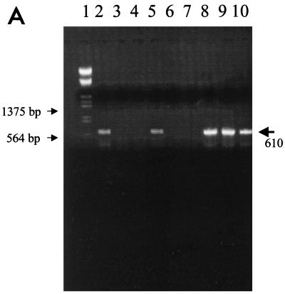
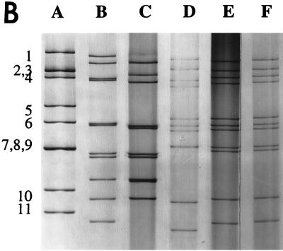
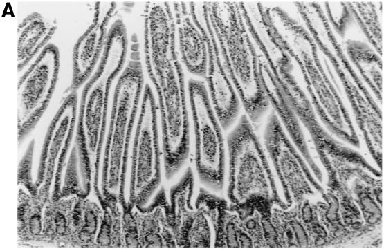
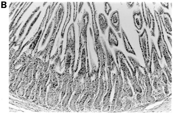
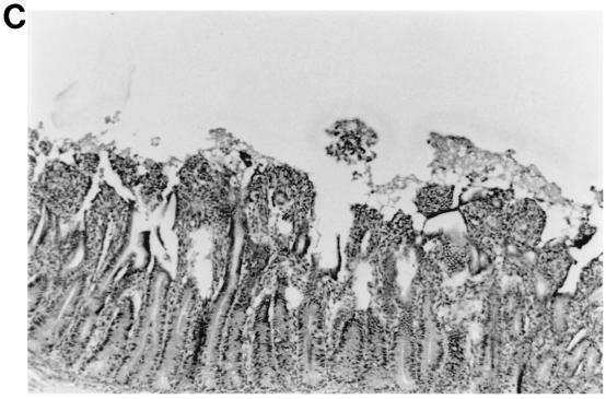
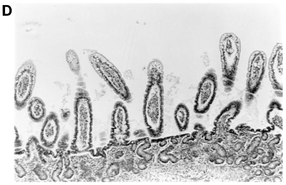
There is serological evidence that bovine group C rotaviruses exist in the United States, but there are no reports of their isolation. Ninety fecal samples from calves with diarrhea, 81 samples from adult cows with diarrhea (winter dysentery), and 20 fecal samples from healthy adult cows were tested for group C rotaviruses by polyacrylamide gel electrophoresis, immune electron microscopy, and reverse transcription-PCR (RT-PCR). Three samples from adult cow diarrhea cases were positive only by RT-PCR, and a group C rotavirus was isolated from a positive sample in monkey kidney (MA104) cells (WD534tc/C). Genetically and serologically, the WD534tc/C strain was more closely related to the Cowden porcine group C strain than to the Shintoku bovine strain. Because the original cow feces also contained a group A rotavirus (detected after passage in cell culture), we hypothesized that such dual-rotavirus infections might play a role in the pathogenesis and host adaptation of rotaviruses. Thus, we examined the pathogenesis of WD534tc/C alone or combined with virulent (IND/A) or attenuated (NCDV/A) bovine group A rotaviruses in gnotobiotic calves. WD534tc/C alone induced diarrhea without (or with limited) virus shedding in inoculated calves (n = 3). In contrast, all calves coinfected with WD534tc/C and IND/A (n = 2) developed diarrhea and shed both viruses, whereas calves coinfected with WD534tc/C and NCDV/A (n = 3) developed diarrhea but did not shed either virus. Infection with WD534tc/C or NCDV/A alone caused only mild villous atrophy (jejunum and/or ileum), whereas dual infection with both viruses induced lesions throughout the small intestine. Although IND/A alone caused villous atrophy, more-widespread small intestinal lesions occurred in calves coinfected with WD534tc/C and IND/A. In conclusion, coinfection of calves with group A rotaviruses enhanced fecal shedding of a bovine group C rotavirus and the extent of histopathological lesions in the small intestines. Thus, our findings suggest a potential novel hypothesis involving dual infections for the adaptation of heterologous rotaviruses to new host species.


Figures

FIG. 1
FIG. 1
(A) The RT-PCR products of reference and field samples detected with primers specific for the partial VP6 gene of group C rotavirus. Lane 1, molecular size markers; lanes 2 to 5, field fecal samples; lane 6, IND/A; lane 7, NCDV/A; lane 8, Cowden/C; lane 9, Shintoku/C; lane 10, WD534tc/C. (B) dsRNA electropherotypes of the reference and WD534tc strains. Lane A, IND/A; lane B, ATI/B; lane C, WD653/B; lane D, Shintoku/C; lane E, Cowden/C; lane F, WD534tc/C. Numbers to the left of lane A designate group A rotavirus dsRNA segments.
FIG. 1
FIG. 1
(A) The RT-PCR products of reference and field samples detected with primers specific for the partial VP6 gene of group C rotavirus. Lane 1, molecular size markers; lanes 2 to 5, field fecal samples; lane 6, IND/A; lane 7, NCDV/A; lane 8, Cowden/C; lane 9, Shintoku/C; lane 10, WD534tc/C. (B) dsRNA electropherotypes of the reference and WD534tc strains. Lane A, IND/A; lane B, ATI/B; lane C, WD653/B; lane D, Shintoku/C; lane E, Cowden/C; lane F, WD534tc/C. Numbers to the left of lane A designate group A rotavirus dsRNA segments.
FIG. 2
FIG. 2
Intestinal lesions in the jejuna of gnotobiotic calves following oronasal inoculation with virus. The calves were mock infected (2 PID) (A) or infected with WD534tc/C (4 PID) (B) or IND/A (3 PID) (C) or coinfected with IND/A and WD534tc/C (2 PID) (D). The slides were stained with hematoxylin and eosin. Note that WD534tc alone caused minimal changes (B), whereas IND/A and coinfection with IND/A and WD534tc/C caused villous atrophy (C and D), vacuolation of absorptive cells (D), and hyperplasia of cryptic cells (C and D) in the jejunum. Magnification, ×50.
FIG. 2
FIG. 2
Intestinal lesions in the jejuna of gnotobiotic calves following oronasal inoculation with virus. The calves were mock infected (2 PID) (A) or infected with WD534tc/C (4 PID) (B) or IND/A (3 PID) (C) or coinfected with IND/A and WD534tc/C (2 PID) (D). The slides were stained with hematoxylin and eosin. Note that WD534tc alone caused minimal changes (B), whereas IND/A and coinfection with IND/A and WD534tc/C caused villous atrophy (C and D), vacuolation of absorptive cells (D), and hyperplasia of cryptic cells (C and D) in the jejunum. Magnification, ×50.

References

    1. Ball J M, Tian P, Zeng C Q, Morris A P, Estes M K. Age dependent diarrhea induced by a rotaviral nonstructural glycoprotein. Science. 1996;272:101–104. - PubMed
    1. Blackhall J, Bellinzoni R, Mattion N, Estes M K, LaTorre J L, Magnusson G. A bovine rotavirus serotype 1: serological characterization of the virus and nucleotide sequence determination of the structural glycoprotein VP7 gene. Virology. 1992;189:833–837. - PubMed
    1. Bohl E H, Saif L J, Theil K W, Agnes A G, Cross R F. Porcine pararotavirus: detection, differentiation from rotavirus, and pathogenesis in gnotobiotic pigs. J Clin Microbiol. 1982;15:312–319. - PMC - PubMed
    1. Bridger J C. Non-group A rotaviruses. In: Kapikian A Z, editor. Virus infections of the gastrontestinal tract. New York, N.Y: Marcel Dekker; 1994. pp. 369–407.
    1. Chang K O, Parwani A V, Smith D, Saif L J. Detection of group B rotaviruses in fecal samples from diarrheic calves and adult cows and characterization of their VP7 genes. J Clin Microbiol. 1997;35:2107–2110. - PMC - PubMed

Publication types

MeSH terms

Associated data