. 1999 Nov;73(11):9413-21.
doi: 10.1128/JVI.73.11.9413-9421.1999.

Requirements for RNA replication of a poliovirus replicon by coxsackievirus B3 RNA polymerase

Affiliations

Requirements for RNA replication of a poliovirus replicon by coxsackievirus B3 RNA polymerase

Y C Bell et al. J Virol. 1999 Nov.

Abstract

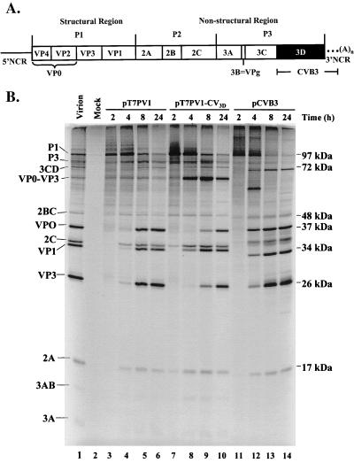
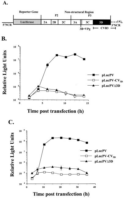
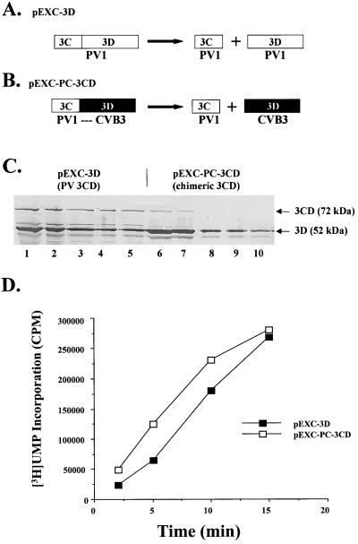
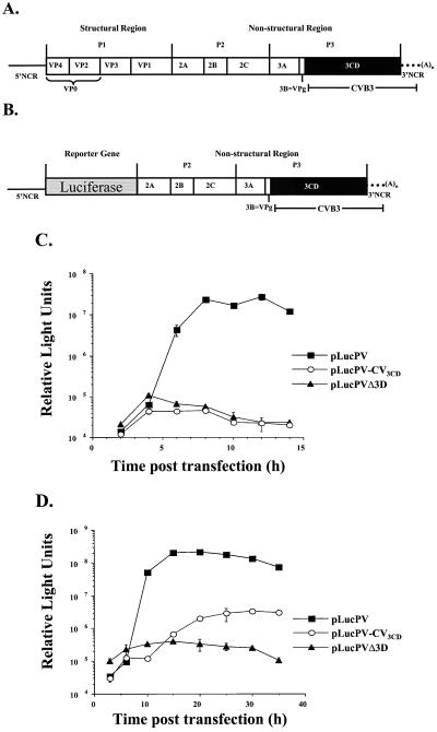
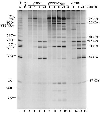
A chimeric poliovirus type 1 (PV1) genome was constructed in which the 3D RNA polymerase (3D(pol)) coding sequences were replaced with those from coxsackievirus B3 (CVB3). No infectious virus was produced from HeLa cells transfected with the chimeric RNA. Processing of the PV1 capsid protein precursor was incomplete, presumably due to inefficient recognition of the P1 protein substrate by the chimeric 3CD proteinase containing CVB3 3D sequences. The ability of the chimeric RNA to replicate in the absence of capsid formation was measured after replacement of the P1 region with a luciferase reporter gene. No RNA synthesis was detected, despite efficient production of enzymatically active 3D(pol) from the 3D portion of the chimeric 3CD. The chimeric 3CD protein was unable to efficiently bind to the cloverleaf-like structure (CL) at the 5' end of PV1 RNA, which has been demonstrated previously to be required for viral RNA synthesis. The CVB3 3CD protein bound the PV1 CL as well as PV1 3CD. An additional chimeric PV1 RNA that contained CVB3 3CD sequences also failed to produce virus after transfection. Since processing of PV1 capsid protein precursors by the CVB3 3CD was again incomplete, a luciferase-containing replicon was also analyzed for RNA replication. The 3CD chimera replicated at 33 degrees C, but not at 37 degrees C. Replacement of the PV1 5'-terminal CL with that of CVB3 did not rescue the temperature-sensitive phenotype. Thus, there is an essential interaction(s) between 3CD and other viral P2 or P3 protein products required for efficient RNA replication which is not fully achieved between proteins from the two different members of the same virus genus.


Figures

FIG. 1
FIG. 1
Capsid processing of PV1 chimeras containing CVB3 3Dpol. (A) Schematic representation of a full-length chimeric genome and viral polyprotein organization are shown. The viral structural proteins are derived from the P1 portion of the genome, and the nonstructural proteins are derived from the P2 and P3 portions of the genome. PV sequences are represented by solid lines and white boxes, whereas CV sequences are represented by the black box (3D coding sequences) and dotted lines (3′ NCR). (B) In vitro translation of PV1, CVB3, and chimeric T7 transcripts. Full-length transcripts from wild-type PV1 cDNA (lanes 3 to 6), chimeric PV1-CVB3 3D cDNA (lanes 7 to 10), and wild-type CVB3 cDNA (lanes 11 to 14) were used to program translation reactions in HeLa S10-ribosomal salt wash extracts. At the indicated times, aliquots were removed, diluted in Laemmli sample buffer, and subjected to electrophoresis on a 12.5% polyacrylamide–SDS gel. Lane 2 contains a mock translation mixture incubated for 8 h with no RNA added. A translation reaction mixture programmed with PV1 virion RNA was loaded in lane 1 as a marker for viral proteins. The positions of viral proteins are indicated to the left and the molecular masses are indicated to the right.
FIG. 2
FIG. 2
Lack of RNA replication of PV chimeras containing CVB3 3Dpol. (A) Schematic representation of a chimeric PV replicon in which the nonstructural genes have been replaced with a luciferase reporter gene. (B) Time course of RNA replication measured by luciferase accumulation following RNA transfection of HeLa cells at 37°C. (C) Time course of RNA replication measured by luciferase accumulation following RNA transfection of HeLa cells at 33°C.
FIG. 3
FIG. 3
(A) Production of PV1 3Dpol and CVB3 3Dpol in E. coli. A schematic of PV1 3CD fusion protein expressed in E. coli is presented. 3CD is processed to generate active PV1 3Cpro and PV1 3Dpol. (B) Schematic of a chimeric 3CD fusion protein expressed in E. coli. The chimeric 3CD is autocleaved to generate PV1 3Cpro and CVB3 3Dpol. (C) Western blot analysis of soluble lysates of 3CD expressed in E. coli. Dilutions of extracts containing wild-type PV1 3CD (lanes 1 to 5) and chimeric 3CD (lanes 6 to 10) were resolved by SDS-PAGE (10% polyacrylamide) and transferred to nitrocellulose membrane for analysis with antiserum raised against PV1 3Dpol. The positions and sizes of viral proteins are indicated to the right. (D) Elongation activities of PV1 3Dpol and CV3 3Dpol generated from autocleavage of 3CD fusion proteins. Portions were taken at the indicated time and measured for UMP incorporation as described in Materials and Methods.
FIG. 4
FIG. 4
Purification and biochemical analysis of His-tagged 3CD proteins expressed in E. coli. His-tagged proteins were expressed in BL21(DE3) cells and affinity purified. (A) Coomassie blue staining of a 12.5% polyacrylamide–SDS gel following electrophoresis of fractions (lane 1, chimeric 3CD; lane 2, CVB3 3CD; lane 3, PV1 3CD) purified on a nickel column. Lane 4 contains marker proteins. (B) Western blot analysis of a gel identical to that shown in panel A. Antiserum raised against PV1 3C was used for immunoblot analysis. (C) RNA electrophoretic mobility shift analysis with an RNA probe representing the PV1 5′ CL structure. PV1 probe was incubated with buffer alone (lane 1), with recombinant PCBP2 alone, or with recombinant PCBP2 and the indicated amounts of 3CD (lanes 3 to 11). RNA-protein complexes were resolved on a 4% native polyacrylamide gel and are indicated to the left of the autoradiograph.
FIG. 5
FIG. 5
RNA replication of a PV-replicon chimera containing CVB3 3CD. (A) A schematic representation of a full-length chimeric PV1 genome structure and viral protein organization similar to that in Fig. 1A is shown. The 3CD coding sequences and 3′ NCR of PV1 genome were replaced with those of CVB3. (B) A schematic representation of a chimeric PV replicon containing CVB3 3CD and 3′ NCR is shown. (C) Time course of RNA replication measured by luciferase accumulation following RNA transfection of HeLa cells at 37°C. (D) Time course of RNA replication measured by luciferase accumulation following RNA transfection of HeLa cells at 33°C.
FIG. 6
FIG. 6
In vitro translation and processing of PV1, CVB3, and chimeric T7 transcripts. Full-length transcripts from wild-type PV1 cDNA (lanes 3 to 6), chimeric PV1-CVB3 3CD cDNA (lanes 7 to 10), and wild-type CVB3 cDNA (lanes 11 to 14) were used to program translation in HeLa S10 extracts (supplemented with a ribosomal salt wash from HeLa cells) in the presence of [35S]methionine. Reactions were analyzed as described in the legend to Fig. 2.
FIG. 7
FIG. 7
Inability of CVB3 5′CL to rescue temperature sensitivity of PV1-CVB3 3CD replicon. (A) Schematic representation of a chimeric PV replicon in which the structural genes have been replaced with a luciferase reporter gene is presented. The first ∼100 5′ nt, 3CD coding region, and 3′ NCR of PV1 sequences were replaced with CVB3 sequences. (B) Time course of RNA replication measured by luciferase accumulation following RNA transfection of HeLa cells at 33°C. In this experiment, pRLuc31-2Cpro, which has a lethal mutation in 2C and does not replicate (25), was used as a negative RNA replication control.

References

    1. Andino R, Rieckhof G E, Achacoso P L, Baltimore D. Poliovirus RNA synthesis utilizes an RNP complex formed around the 5′-end of viral RNA. EMBO J. 1993;12:3587–3598. - PMC - PubMed
    1. Andino R, Rieckhof G E, Baltimore D. A functional ribonucleoprotein complex forms around the 5′ end of poliovirus RNA. Cell. 1990;63:369–380. - PubMed
    1. Bell, Y. C., J. H. C. Nguyen, and B. L. Semler. Unpublished observations.
    1. Bienz K, Egger D, Pasamontes L. Association of polioviral proteins of the P2 genomic region with the viral replication complex and virus-induced membrane synthesis as visualized by electron microscopic immunocytochemistry and autoradiography. Virology. 1987;160:220–226. - PubMed
    1. Blair W S, Nguyen J H, Parsley T B, Semler B L. Mutations in the poliovirus 3CD proteinase S1-specificity pocket affect substrate recognition and RNA binding. Virology. 1996;218:1–13. - PubMed

Publication types

MeSH terms

LinkOut - more resources