. 1999 Nov;73(11):9446-55.
doi: 10.1128/JVI.73.11.9446-9455.1999.

Repeated delivery of adeno-associated virus vectors to the rabbit airway

Affiliations

Repeated delivery of adeno-associated virus vectors to the rabbit airway

S E Beck et al. J Virol. 1999 Nov.

Abstract

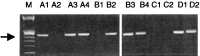
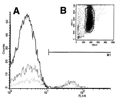
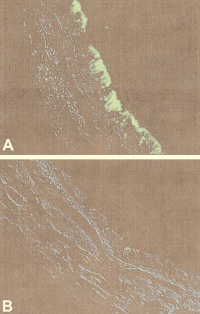
Efficient local expression from recombinant adeno-associated virus (rAAV)-cystic fibrosis (CF) transmembrane conductance regulator (CFTR) vectors has been observed in the airways of rabbits and monkeys for up to 6 months following a single bronchoscopic delivery. However, it is likely that repeated administrations of rAAV vectors will be necessary for sustained correction of the CF defect in the airways. The current study was designed to test the feasibility of repeated airway delivery of rAAV vectors in the rabbit lung. After two doses of rAAV-CFTR to the airways, rabbits generated high titers of serum anti-AAV neutralizing antibodies. Rabbits then received a third dose of a rAAV vector containing the green fluorescent protein (GFP) reporter gene packaged in either AAV serotype 2 (AAV2) or serotype 3 (AAV3) capsids. Each dose consisted of 1 ml containing 5 x 10(9) DNase-resistant particles of rAAV vector, having no detectable replication-competent AAV or adenovirus. Three weeks later, GFP expression was observed in airway epithelial cells despite high anti-AAV neutralizing titers at the time of delivery. There was no significant difference in the efficiency of DNA transfer or expression between the rAAV3 and rAAV2 groups. No significant inflammatory responses to either repeated airway exposure to rAAV2-CFTR vectors or to GFP expression were observed. These experiments demonstrate that serum anti-AAV neutralizing antibody titers do not predict airway neutralization in vivo and that repeated airway delivery rAAV allows for safe and effective gene transfer.


Figures

FIG. 1
FIG. 1
Study design. rAAV2-CFTR vector was instilled into the RLL, tracheas, and noses of NZW rabbits (n = 31) at week 0, and a second dose was given at week 3. The rabbits were then divided into three groups and received either rAAV3-GFP (n = 10), rAAV2-GFP (n = 11), or rAAV2-CFTR (n = 10) to both the RLL and LLL at week 17. Animals that received the heterologous transgene (GFP) vectors were sacrificed at week 20; rabbits that received the homologous transgene (CFTR) as the third dose were sacrificed at week 21. X indicates the study week during which the indicated intervention was performed. *, the GFP groups underwent cytologic brushings of the bronchial mucosa prior to sacrifice. BAL fluid (for total cell counts and neutralizing antibody) and serum (for neutralizing antibody) were obtained immediately prior to each delivery and at sacrifice. Additional serum was obtained at week 6 for neutralizing antibody assay. n indicates number of rabbits in each group.
FIG. 2
FIG. 2
Map of vector and packaging constructs. The DNA contained in the rAAV-CFTR vector contains full-length hCFTR cDNA inserted between the ITRs of AAV2 (ITR-2). This vector was packaged in AAV2 capsids by using a cell line that expresses Cap protein from AAV2 (Cap-2) and is referred to as rAAV2-CFTR in this study. The DNA contained in the rAAV-GFP vectors, TR-UF5, contains the cytomegalovirus (CMV)-driven Neor gene and humanized GFP cDNA reporter gene cassette inserted between the ITRs of AAV2 (ITR-2). rAAV-GFP vector DNA was packaged in AAV2 capsids by using the packaging construct pRS5 and is referred to as rAAV2-GFP vector. rAAV-GFP vector DNA was also packaged in AAV3 capsids by using the construct pSB-Cap3.6, which contains the Cap gene from AAV3 (Cap-3) in place of Cap from AAV2 (Cap-2) in the packaging construct (see text for explanation), and is referred to as rAAV3-GFP vector. TK, thymidine kinase; LTR, long terminal repeat.
FIG. 3
FIG. 3
GFP expression in 293 cells following infection with 104 DRP of rAAV2-GFP vector (B) or rAAV3-GFP vector (C) per cell. (A) Uninfected 293 cells.
FIG. 4
FIG. 4
Serum neutralizing AAV antibody response. The left-hand side shows the baseline titer at week 0, the response 3 weeks after the first dose (week 3), and the responses 3 and 14 weeks after the second dose (week 6 and week 17, respectively). The right-hand side shows the responses 3 weeks after administration of the third dose of either rAAV3-GFP (A) or rAAV2-GFP (B) and 4 weeks after readministration of a dose of rAAV2-CFTR (C). Bars represent median values, shaded boxes represent 2 quartiles of values, whiskers represent ranges of values, ∗ indicates significance at P < 0.001, and n/s (not significant) indicates significance at P > 0.05. Note that the titer is plotted on a log2 scale. n values are as indicated in Fig. 1.
FIG. 5
FIG. 5
Ethidium bromide-stained 1.5% agarose gel electrophoresis analysis of vector-specific DNA amplified from the RLL (even numbers) or LLL (odd numbers) of rAAV3-GFP-treated (A1 to A4), rAAV2-GFP-treated (B1 to B4), and vehicle-treated (C1 to C2) animals. pTR-UF5 plasmid dilutions D1 (10−14 g) and D2 (10−17 g) in a background of 200 ng of untreated rabbit lung DNA show the sensitivity of the PCR. M is the 100-bp marker; the arrow marks expected 355-bp PCR product.
FIG. 6
FIG. 6
FACS analysis of brushed bronchial epithelial cells taken 3 weeks after the third dose of AAV vector. (A) Representative histogram of brushed bronchial epithelial cells from the RLL and LLL (dotted lines) of an AAV3-GFP-treated rabbit showing 45 and 25% of cells with characteristic GFP fluorescence compared to an untreated control animal (solid line) showing less than 0.5% fluorescing cells. (B) Forward/side scatter profile of epithelial cells gated for analysis.
FIG. 7
FIG. 7
GFP expression in rabbit bronchial epithelium after the third dose of rAAV vector. Proximal RLLs were sectioned at the level of segmental branching, fixed in 10% formalin, and analyzed by GFP-specific fluorescent microscopy. (A) GFP in the cytoplasm of ciliated columnar epithelial cells in the RLL of an AAV3-GFP-treated rabbit; (B) vehicle-treated rabbit airway showing minimal background autofluorescence. Original magnification, ×400. Sections were examined under differential interference contrast, captured, colored blue, and overlaid onto the fluorescent image to show underlying airway architecture.
FIG. 8
FIG. 8
Inflammatory profile of BAL fluid before and after treatment with rAAV vectors. (A) IL-8 in rabbit BAL fluid after three doses of rAAV vector and in BAL fluid from Ad vector-infected rabbits (striped bar at the bottom; n = 4); (B) total BAL fluid cell counts; (C) percentages of macrophages in BAL fluid; (D) percentages of lymphocytes in BAL fluid; (E) percentages of eosinophils (Eos) in BAL fluid. Designations for repeatedly dosed groups (diagonally hatched bars; n = 10 to 11 in each group), single-dose controls (stippled bars; n = 2 in each group), and pre-first dose (open bar at top; n = 31) apply to all panels. Bars represent means, whiskers represent standard errors of the means, and ∗ indicates significance at P < 0.05.
FIG. 9
FIG. 9
Histopathology of rabbit lung. Five-micrometer sections of formalin-fixed lung, stained with hematoxylin and eosin, from each animal in the study were examined as described in Materials and Methods. (A) LLL of the thrice-dosed AAV2-CFTR group (received only one local dose of rAAV2-CFTR). Note BALT in the submucosa of this cartilaginous airway and the intact alveolar and airway architecture. This finding was typical in all treated and untreated rabbits in the study. Original magnification, ×40. (B) RLL after three instillations of rAAV2-CFTR. Note minimal lymphocytic infiltration in the submucosa of a large airway with preservation of airway architecture. This degree of lymphocytic infiltration was common in treated and untreated rabbits. Original magnification, ×100. (C) RLL of a single-dose vehicle-treated control rabbit showing intact epithelium, normal alveolar architecture, and BALT (upper left-hand corner), typical of all rAAV-treated vehicle-treated, and untreated rabbits in the study. Original magnification, ×40. (D) For comparison, a rabbit airway 3 days after instillation of 107 PFU of an Ad vector, showing inflammatory cell disruption of the epithelium and submucosa of a large airway. This was not seen in any rAAV- or vehicle-treated rabbit in the study. Original magnification, ×40. (E) Same animal as in panel A, showing small focal aggregate of lymphocytes and eosinophils around a bronchiole and structural integrity of alveolar air spaces, epithelium, and submucosa. This finding was typical of all treated and untreated rabbits in the study. Original magnification, ×100. (F) Same animal as in panel B, showing structural integrity of epithelium and basement membrane, with occasional lymphocytes and eosinophils, and lack of neutrophils in submucosa. This finding was typical of all treated and untreated rabbits in the study. Original magnification, ×200. (G) Same as animal as in panel C, showing integrity of epithelium and basement membrane, typical of all rAAV- and vehicle-treated rabbits in the study. Original magnification, ×200. (H) Same animal as panel D, showing inflammatory cell infiltration. Note neutrophils and eosinophils invading the columnar epithelial cells, which were not seen in any rAAV- or vehicle-treated rabbit in the study. Original magnification, ×100.

References

    1. Afione S A, Conrad C K, Kearns W G, Chunduru S, Adams R, Reynolds T C, Guggino W B, Cutting G R, Carter B J, Flotte T R. In vivo model of adeno-associated virus vector persistence and rescue. J Virol. 1996;70:3235–3241. - PMC - PubMed
    1. Ayers M M, Jeffery P K. Proliferation and differentiation in mammalian airway epithelium. Eur Respir J. 1988;1:58–80. - PubMed
    1. Balcklow N R. Adeno-associated viruses of humans. In: Pattison J R, editor. Parvoviruses and human disease. Boca Raton, Fla: CRC Press, Inc.; 1988. pp. 165–174.
    1. Bantel-Schaal U, zur Hausen H H. Characterization of the DNA of a defective human parvovirus isolated from a genital site. Virology. 1984;134:52–63. - PubMed
    1. Bartlett J S, Samulski R J, McCown T J. Selective and rapid uptake of adeno-associated virus type 2 in brain. Hum Gene Ther. 1998;9:1181–1186. - PubMed

Publication types

MeSH terms

Substances

LinkOut - more resources