. 1999 Nov;73(11):9532-43.
doi: 10.1128/JVI.73.11.9532-9543.1999.

Mutant cells selected during persistent reovirus infection do not express mature cathepsin L and do not support reovirus disassembly

Affiliations

Mutant cells selected during persistent reovirus infection do not express mature cathepsin L and do not support reovirus disassembly

G S Baer et al. J Virol. 1999 Nov.

Abstract

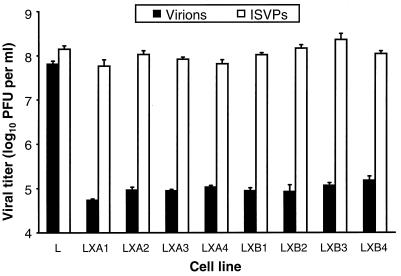
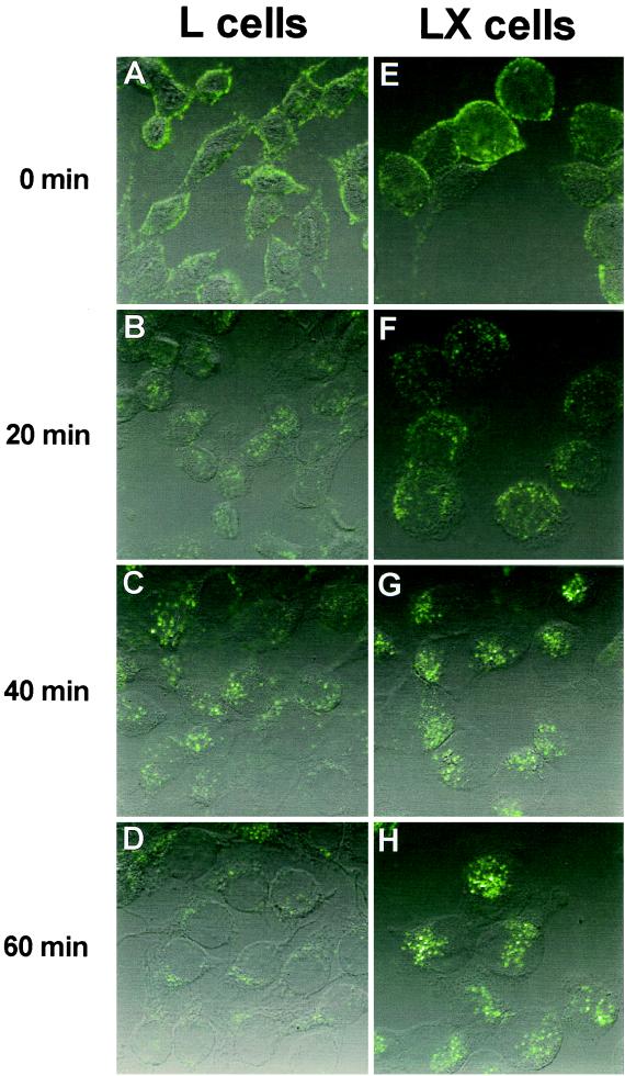
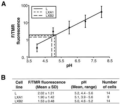
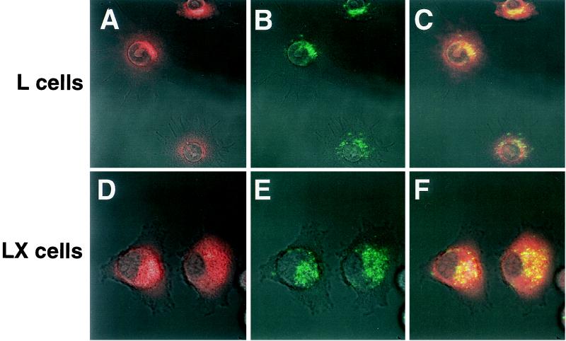
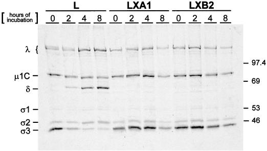
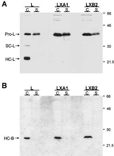
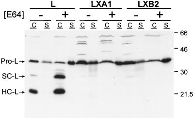
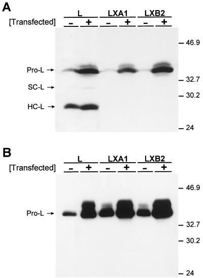
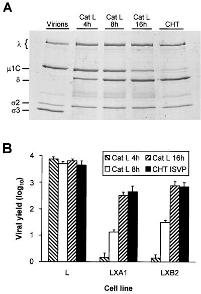
Persistent reovirus infections of murine L929 cells select cellular mutations that inhibit viral disassembly within the endocytic pathway. Mutant cells support reovirus growth when infection is initiated with infectious subvirion particles (ISVPs), which are intermediates in reovirus disassembly formed following proteolysis of viral outer-capsid proteins. However, mutant cells do not support growth of virions, indicating that these cells have a defect in virion-to-ISVP processing. To better understand mechanisms by which viruses use the endocytic pathway to enter cells, we defined steps in reovirus replication blocked in mutant cells selected during persistent infection. Subcellular localization of reovirus after adsorption to parental and mutant cells was assessed using confocal microscopy and virions conjugated to a fluorescent probe. Parental and mutant cells did not differ in the capacity to internalize virions or distribute them to perinuclear compartments. Using pH-sensitive probes, the intravesicular pH was determined and found to be equivalent in parental and mutant cells. In both cell types, virions localized to acidified intracellular organelles. The capacity of parental and mutant cells to support proteolysis of reovirus virions was assessed by monitoring the appearance of disassembly intermediates following adsorption of radiolabeled viral particles. Within 2 h after adsorption to parental cells, proteolysis of viral outer-capsid proteins was observed, consistent with formation of ISVPs. However, in mutant cells, no proteolysis of viral proteins was detected up to 8 h postadsorption. Since treatment of cells with E64, an inhibitor of cysteine-containing proteases, blocks reovirus disassembly, we used immunoblot analysis to assess the expression of cathepsin L, a lysosomal cysteine protease. In contrast to parental cells, mutant cells did not express the mature, proteolytically active form of the enzyme. The defect in cathepsin L maturation was not associated with mutations in procathepsin L mRNA, was not complemented by procathepsin L overexpression, and did not affect the maturation of cathepsin B, another lysosomal cysteine protease. These findings indicate that persistent reovirus infections select cellular mutations that affect the maturation of cathepsin L and suggest that alterations in the expression of lysosomal proteases can modulate viral cytopathicity.


Figures

FIG. 1
FIG. 1
Viral titers in parental L cells and independent mutant LX cell clones after infection by virions and ISVPs. Monolayers of parental L cells and eight independent mutant LX-cell clones (5 × 10⁵ cells) were infected with either virions or ISVPs of reovirus T1L at an MOI of 2 PFU per cell. After a 1-h adsorption period, the inoculum was removed, fresh medium was added, and the cells were incubated at 37°C for 24 h. Cells were frozen and thawed twice, and viral titers in cell lysates were determined by plaque assay using L-cell monolayers. The results are presented as mean viral titers for four independent experiments. Error bars indicate standard deviations of the means.
FIG. 2
FIG. 2
Ultrastructural morphologies of uninfected, persistently infected, and cured L cells. (A) Uninfected parental L cells. (B, C, and D) Persistently infected LDG cells. Note in panel B the large inclusion of virions (arrow). (D) Increased magnification of electron-dense vesicles from panel C. (E and F) Cured LXA1 cells. Note the presence of electron-dense vesicles in cured cells. Bars, 5 μm.
FIG. 3
FIG. 3
Uptake of reovirus virions following adsorption to parental L cells and mutant LXA1 cells. Monolayers of L cells (A to D) and LXA1 cells (E to H) were adsorbed with 10,000 particles per cell of Cy3-conjugated virions of reovirus strain T1L at 4°C for 45 min. Following removal of unbound virus, cells were incubated at 37°C for 0 (A and E), 20 (B and F), 40 (C and G), or 60 (D and H) min and then fixed. Cells were visualized by confocal fluorescence microscopy.
FIG. 4
FIG. 4
Determination of intravesicular pHs of parental L cells and mutant LX cells. (A) Standard curve correlating the F/TMR fluorescence ratio with pH. Cells were incubated with double-labeled fluorescein-tetramethylrhodamine dextran (0.1% [wt/vol]) at 37°C for 16 h. Cells were fixed, equilibrated for 1 h with buffers of known pH (0.1 M sodium acetate [pH 4.0 and pH 5.0], 0.1 M sodium phosphate [pH 6.0 and pH 7.0], and 0.1 M Tris [pH 8.0]), and visualized by confocal fluorescence microscopy. Fluorescence intensities for fluorescein and tetramethylrhodamine were determined for identical groups of 15 to 20 cells at each pH standard. (B) Intravesicular pHs of parental L cells and mutant LX cells determined by calculating the mean F/TMR fluorescence ratio for each cell type and extrapolating from the standard curve, as indicated in panel A.
FIG. 5
FIG. 5
Colocalization of reovirus virions to acidic compartments in parental L cells and mutant LX cells. Virions of reovirus strain T1L conjugated to Cy5 were adsorbed to monolayers of parental L cells and mutant LXB2 cells (10,000 particles per cell) at 4°C for 45 min. Following removal of unbound virus, cells were incubated in medium containing 3 μM LysoSensor Green probe at 37°C for 1.5 h. Cells were washed and visualized by confocal fluorescence microscopy. Acidic compartments are indicated in red (A and D). Virions are indicated in green (B and E). In the merged image, yellow indicates colocalization of virions and acidic compartments (C and F).
FIG. 6
FIG. 6
Electrophoretic analysis of reovirus structural proteins after infection of parental L cells and mutant LX cells. Monolayers of parental L cells and mutant LXA1 and LXB2 cells (107) were adsorbed with 10,000 particles per cell of purified 35S-labeled virions of reovirus strain T1L. After 1 h of adsorption at 4°C, the inoculum was removed, fresh medium was added, and the cells were incubated at 37°C for the indicated intervals. Cells then were lysed and extracted with freon. Virus particles contained in supernatants were pelleted by ultracentrifugation and solubilized in sample buffer. Equal volumes of samples were loaded into wells of a 10% polyacrylamide gel. After electrophoresis, the gel was prepared for autoradiography and exposed to film. Viral proteins are labeled, and molecular mass standards (in kilodaltons) are indicated.
FIG. 7
FIG. 7
Immunoblot analysis of cathepsin L and cathepsin B expression in cytoplasmic extracts and culture supernatants from parental L cells and mutant LX cells. Monolayers of parental L cells and mutant LXA1 and LXB2 cells (107) were incubated in serum-free medium for 6 h. Cellular proteins (C), normalized for protein content, and secreted proteins (S), normalized for cell number, were resolved in a 12% polyacrylamide gel, electroblotted onto a nitrocellulose membrane, and immunoblotted with rabbit antisera raised against either murine cathepsin L (A) or human cathepsin B (B). Horseradish peroxidase-conjugated anti-rabbit immunoglobulin G antiserum was used as secondary antibody, and proteins were visualized by chemiluminescence. Bands corresponding to procathepsin L (Pro-L), the single-chain form of cathepsin L (SC-L), the heavy-chain form of cathepsin L (HC-L), and the heavy-chain form of cathepsin B (HC-B) are indicated. Molecular mass standards (in kilodaltons) are shown.
FIG. 8
FIG. 8
Effect of protease inhibitor E64 on steady-state levels and secretion of cathepsin L from parental L cells and mutant LX cells. Monolayers of parental L cells and mutant LXA1 and LXB2 cells (107) were incubated at 37°C for 18 h in the presence or absence of 200 μM E64. Cellular proteins (C), normalized for protein content, and secreted proteins (S), normalized for cell number, were resolved in a 12% polyacrylamide gel, electroblotted onto a nitrocellulose membrane, and immunoblotted with rabbit anti-cathepsin L antiserum. Horseradish peroxidase-conjugated anti-rabbit immunoglobulin G antiserum was used as secondary antibody, and proteins were visualized by chemiluminescence. Bands corresponding to procathepsin L (Pro-L) and the single-chain (SC-L) and heavy-chain (HC-L) forms of cathepsin L are indicated. Molecular mass standards (in kilodaltons) are shown.
FIG. 9
FIG. 9
Effect of procathepsin L overexpression on steady-state levels and secretion of cathepsin L from parental L cells and mutant LX cells. Monolayers of parental L cells and mutant LXA1 and LXB2 cells at approximately 80% confluence were transfected with a plasmid encoding murine procathepsin L and incubated at 37°C for 72 h. Equal amounts of cellular proteins (A) and equal amounts of secreted proteins (B) were resolved in 12% polyacrylamide gels, electroblotted onto nitrocellulose membranes, immunoblotted with rabbit anti-cathepsin L antiserum, and visualized by chemiluminescence. Bands corresponding to procathepsin L (Pro-L) and the single-chain (SC-L) and heavy-chain (HC-L) forms of cathepsin L are indicated. Molecular mass standards (in kilodaltons) are shown.
FIG. 10
FIG. 10
Treatment of reovirus virions with purified cathepsin L. (A) Electrophoretic analysis of viral structural proteins of reovirus virions after treatment with either cathepsin L (Cat L) or chymotrypsin (CHT). Purified virions of reovirus strain T1L were treated with either human cathepsin L (pH 5.0) at 37°C for the indicated intervals or bovine α-chymotrypsin (pH 7.4) at 37°C for 2 h. Virions also were incubated at 37°C in virion storage buffer adjusted to pH 5.0 for 16 h. Equal numbers of virus particles were loaded into wells of a 10% polyacrylamide gel. After electrophoresis, the gel was stained with Coomassie blue. Viral proteins are labeled. (B) Growth of virions treated with cathepsin L and ISVPs generated by chymotrypsin in parental L cells and mutant LX cells. Monolayers of cells (4 × 10⁵ cells) were infected with either T1L virions treated with cathepsin L for 4 (Cat L 4 h), 8 (Cat L 8 h), or 16 (Cat L 16 h) h or ISVPs generated by treatment of T1L virions with chymotrypsin for 2 h (CHT ISVP) at an MOI of 2 PFU per cell. After a 1-h adsorption period, the inoculum was removed, fresh medium was added, and cells were incubated at 37°C for either 0 or 24 h. Cells were frozen and thawed twice, and viral titers in cell lysates were determined by plaque assay using L-cell monolayers. The results are presented as mean viral yields, calculated by dividing viral titers at 24 h by viral titers at 0 h, for three independent experiments. Error bars indicate standard deviations of the means.

References

    1. Ahmed R, Canning W M, Kauffman R S, Sharpe A H, Hallum J V, Fields B N. Role of the host cell in persistent viral infection: coevolution of L cells and reovirus during persistent infection. Cell. 1981;25:325–332. - PubMed
    1. Ahmed R, Chakraborty P R, Graham A F, Ramig R F, Fields B N. Genetic variation during persistent reovirus infection: presence of extragenically suppressed temperature-sensitive lesions in wild-type virus isolated from persistently infected L cells. J Virol. 1980;34:383–389. - PMC - PubMed
    1. Ahmed R, Fields B N. Role of the S4 gene in the establishment of persistent reovirus infection in L cells. Cell. 1982;28:605–612. - PubMed
    1. Ahmed R, Graham A F. Persistent infections in L cells with temperature-sensitive mutants of reovirus. J Virol. 1977;23:250–262. - PMC - PubMed
    1. Ahmed R, Kauffman R S, Fields B N. Genetic variation during persistent reovirus infection: isolation of cold-sensitive and temperature-sensitive mutants from persistently infected L cells. Virology. 1983;131:71–78. - PubMed

Publication types

MeSH terms

Associated data

LinkOut - more resources