. 1999 Oct 15;19(20):8818-29.
doi: 10.1523/JNEUROSCI.19-20-08818.1999.

Identification of a cis-acting dendritic targeting element in MAP2 mRNAs

Affiliations

Identification of a cis-acting dendritic targeting element in MAP2 mRNAs

A Blichenberg et al. J Neurosci. .

Abstract

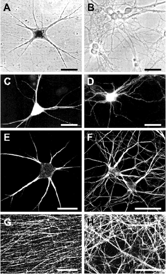
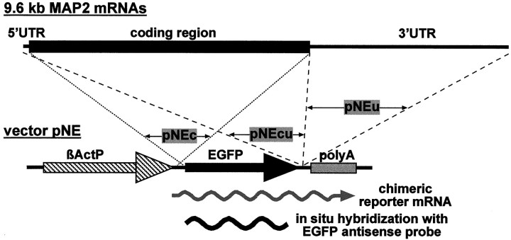
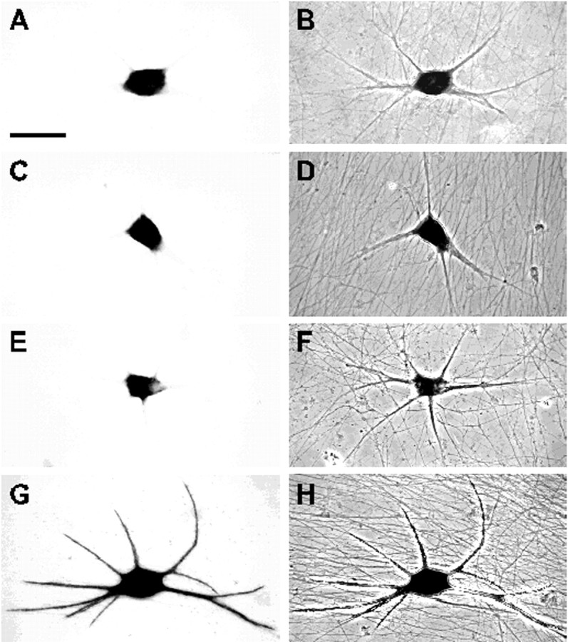
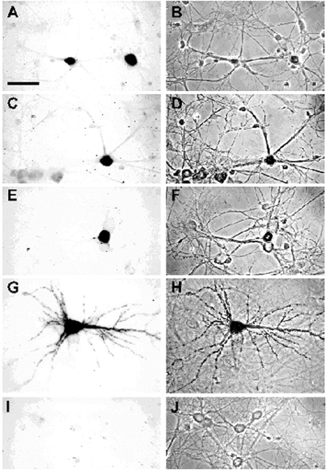
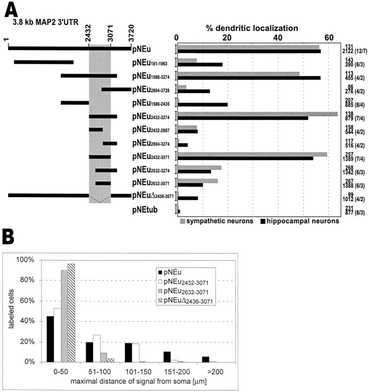
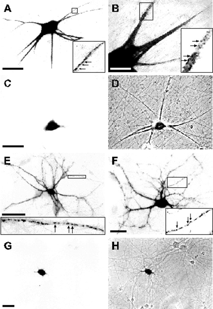
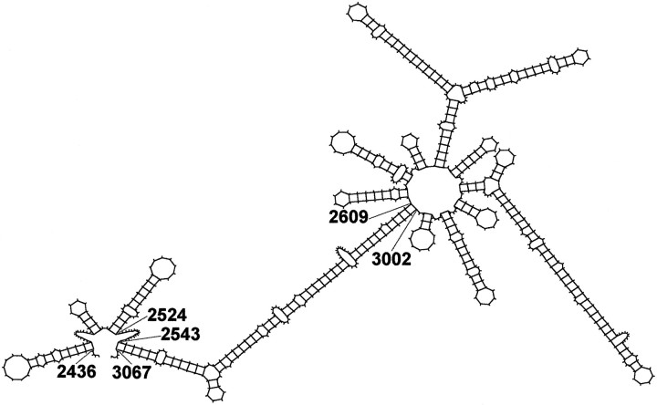
In neurons, a limited number of mRNAs have been identified in dendritic processes, whereas other transcripts are restricted to the cell soma. Here we have investigated the molecular mechanisms underlying extrasomatic localization of mRNAs encoding microtubule-associated protein 2 (MAP2) in primary neuronal cultures. Vectors expressing recombinant mRNAs were introduced into hippocampal and sympathetic neurons using DNA transfection and microinjection protocols, respectively. Chimeric mRNAs containing the entire 3' untranslated region of MAP2 transcripts fused to a nondendritic reporter mRNA are detected in dendrites. In contrast, RNAs containing MAP2 coding and 5' untranslated regions or tubulin sequences are restricted to the cell soma. Moreover, 640 nucleotides from the MAP2 3' untranslated region (UTR) are both sufficient and essential for extrasomatic localization of chimeric mRNAs in hippocampal and sympathetic neurons. Thus, a cis-acting dendritic targeting element that is effective in two distinct neuronal cell types is contained in the 3' UTR of MAP2 transcripts. The observation of RNA granules in dendrites implies that extrasomatic transcripts seem to assemble into multimolecular complexes that may function as transport units.


Figures

Fig. 1.
Fig. 1.
Polar morphology and protein compartmentalization in primary neurons. Phase-contrast (A, B) and fluorescent (C–H) micrographs of cultured sympathetic (A, C, E, G) and hippocampal neurons (B, D, F, H) after 2 weeks in culture. Phase-contrast images shown in A and Breveal an elaborated neuronal network present in both primary cell cultures. C, D, Neurons were microinjected and transfected, respectively, with pNE vector DNA leading to the expression of EGFP. The recombinant protein was either directly visualized by autofluorescence (D) or detected through immunocytochemistry with an anti-EGFP antibody and a Cy2-coupled secondary antibody (C).EH, Neurons immunostained with mouse monoclonal antibodies against the cytoskeletal proteins MAP2 (E, F) and tau (G, H) and secondary goat anti-mouse antibodies coupled to Cy3. Neuronal somata and dendrites are intensely stained with anti-MAP2 antibodies (E, F). Dendrites taper over their length and often form secondary branches. In hippocampal cultures, dendrites are longer relative to the cell body diameter and branch more frequently than processes of sympathetic neurons. G, H,Anti-tau antibodies primarily label the finely meshed axonal network consisting of thin processes that course in and out of the field of view. Processes often assemble to thicker axon bundles.A–D represent scanned prints of pictures taken with a conventional Leitz fluorescent microscope. Micrographs shown inE–H were captured with a laser-scanning microscope. Scale bars, 50 μm.
Fig. 2.
Fig. 2.
Schematic representation of the expression and detection of recombinant transcripts in primary neurons to identifycis-acting dendritic targeting signals in MAP2 mRNAs. Different MAP2 cDNA fragments are inserted adjacent to the EGFP cDNA in vector pNE. From pNEc a MAP2/EGFP fusion protein encoding mRNA is transcribed, whereas in pNEcu and pNEu MAP2 sequences are inserted downstream of the EGFP stop codon. Subcellular localization of chimeric transcripts in primary neurons is determined by nonradioactivein situ hybridizations with a digoxygenin-labeled RNA probe complementary to EGFP sequences. βActP, β-actin promoter; EGFP, enhanced green fluorescent protein; polyA, polyadenylation signal.
Fig. 3.
Fig. 3.
In situ hybridization analysis of the subcellular distribution of chimeric mRNAs in sympathetic neurons after vector DNA injection into the nucleus. Bright-field (A, C, E, G) and phase-contrast (B, D, F, H) micrographs of cells injected with pNEtub (A, B), pNEcu (C, D), pNEc (E, F), and pNEu (G, H). In neurons shown in A–H, reporter mRNAs were immunocytochemically detected using a digoxigenin-labeled antisense EGFP probe and an alkaline phosphatase-conjugated sheep anti-digoxigenin antibody. After the addition of nitro blue tetrazolium and 5-bromo-4-chloro-3-indolyl phosphate, a dark-colored reaction product was detectable. Chimeric RNAs containing α-tubulin sequences (A, B) or the MAP2 coding region (C, D, E, F) are restricted to cell somata, whereas transcripts carrying the entire 3′ UTR of MAP2 mRNAs are found in dendrites (G, H). Images were collected with a video camera connected to a Leitz microscope. Scale bar, 50 μm.
Fig. 4.
Fig. 4.
Detection of chimeric transcripts in hippocampal neurons after transfection with vector DNA. Bright-field (A, C, E, G, I) and phase-contrast (B, D, F, H, J) micrographs of neurons transfected with pNEtub (A, B, I, J), pNEcu (C, D), pNEc (E, F), and pNEu (G, H) are shown. In AH, neurons were hybridized with a digoxigenin-labeled antisense EGFP probe. After incubation with alkaline phosphatase-conjugated sheep anti-digoxigenin antibody, the addition of nitro blue tetrazolium and 5-bromo-4-chloro-3-indolyl phosphate resulted in a dark-colored reaction product in a small fraction of the cells. Hybridization with a sense EGFP probe never resulted in significant cell staining (I, J). Recombinant transcripts with sequences of the MAP2 coding region (C–F) or α-tubulin mRNA (A, B) are only detected in the cell soma. In contrast, chimeric mRNAs possessing the entire 3′ UTR of MAP2 transcripts are present in dendrites (G, H). Cells were photographed with a video camera attached to a Leitz microscope. Scale bar, 50 μm.
Fig. 5.
Fig. 5.
Subcellular distribution of chimeric transcripts in sympathetic and hippocampal neurons. In the left panel, the molecular structure of 9.6 kb MAP2 transcripts is schematically indicated with black bars andlines representing coding and noncoding regions, respectively. Below, MAP2 sequences that are present in recombinant mRNAs derived from the corresponding vectors are indicated asheavy black bars. From pNEc, a MAP2/EGFP fusion protein is synthesized (indicated by a black arrow), whereas the MAP2 coding region in pNEcu transcripts is not translated into a protein. The right panel lists the relative amount of cells showing dendritic localization of chimeric transcripts in sympathetic and hippocampal neurons and the total number of evaluated cells per construct. In both types of transgenic primary neurons, only recombinant mRNAs containing the MAP2 3′ UTR are efficiently localized to dendrites.
Fig. 6.
Fig. 6.
A, Identification of a 640 nucleotide cis-acting DTE in the 3′ UTR of MAP2 transcripts. In the left panel, regions of the MAP2 3′ UTR included in chimeric mRNAs transcribed from the corresponding vectors are shown as heavy black bars. In theright panel, the percentage of primary neurons exhibiting dendritic mRNA localization patterns and the total number of analyzed cells are shown. In parentheses, the total number of analyzed coverslips/independent transfections of hippocampal neurons are shown. Only chimeric transcripts containing a 640-nucleotide-long sequence highlighted in gray are detected with high frequency in dendrites of sympathetic and hippocampal neurons. Deletion of this targeting element omits dendritic localization. B, Dendritic mRNA localization patterns of several hybrid mRNAs. The histogram displays the relative amount of cells that show their distal-most in situ staining in the indicated ranges of distance from the soma. For each construct, 200 cells from two independent transfections of hippocampal neurons were analyzed. In cells that exhibit a low frequency of dendritic mRNA localization (pNEu2632–3071 and pNEuΔ2436–3071 transfected cells), recombinant transcripts on average traveled less far into the cell processes than in neurons with a high percentage of dendritic labeling (pNEu and pNEu2432–3071).
Fig. 7.
Fig. 7.
In situ hybridization analysis of the subcellular distribution of chimeric mRNAs in sympathetic (A–D) and hippocampal neurons (E–H). Bright-field (A–C, E–G) and phase-contrast (D, H) micrographs of cells expressing recombinant transcripts from pNEu2432–3071(A, B, E, F) and pNEuΔ2436–3071(C, D, G, H) are shown. The 640 nucleotide DTE in the MAP2 3′ UTR is sufficient to mediate extrasomatic mRNA localization in both neuronal cell types (A, B, E, F). Higher magnification images shown as insets depict the particulate nature of the color-reaction product of the nonradioactivein situ hybridization in dendrites (arrows). Deletion of the DTE from the 3′ UTR omits the dendritic targeting capacity (C, D, G, H). Images were captured either with a video camera (A–D) or on conventional film (E–H). Cand G are bright-field images of areas shown as phase-contrast micrographs in D and H, respectively. Scale bars: A, C,E, 50 μm; B,F, G, 20 μm.
Fig. 8.
Fig. 8.
Predicted secondary structure of the dendritic targeting element in rat MAP2 mRNAs. The structure was determined using the mfold program 2.3 developed by Zuker (1989) included in the Wisconsin Package (Genetics Computer Group, Madison, WI). The free energy of the structure shown is −160.7 kcal/mol.

References

    1. Ainger K, Avossa D, Morgan F, Hill SJ, Barry C, Barbarese E, Carson JH. Transport and localization of exogenous myelin basic protein mRNA microinjected into oligodendrocytes. J Cell Biol. 1993;123:431–441. - PMC - PubMed
    1. Bassell G, Singer RH. mRNA and cytoskeletal filaments. Curr Opin Cell Biol. 1997;9:109–115. - PubMed
    1. Bassell GJ, Zhang H, Byrd AL, Femino AM, Singer RH, Taneja KL, Lifshitz LM, Herman IM, Kosik KS. Sorting of β-actin mRNA and protein to neurites and growth cones in culture. J Neurosci. 1998;18:251–265. - PMC - PubMed
    1. Bruckenstein DA, Lein PJ, Higgins D, Fremeau RT., Jr Distinct spatial localization of specific mRNAs in cultured sympathetic neurons. Neuron. 1990;5:809–819. - PubMed
    1. Burgin KE, Waxham MN, Rickling S, Westgate SA, Mobley WC, Kelly PT. In situ hybridization histochemistry of Ca2+/calmodulin-dependent protein kinase in developing rat brain. J Neurosci. 1990;10:1788–1798. - PMC - PubMed

Publication types

Substances

LinkOut - more resources