

Review
doi: 10.1111/j.1469-7793.1999.00033.x.
Proteins involved in synaptic vesicle trafficking
Affiliations
- PMID: 10517798
- PMCID: PMC2269560
- DOI: 10.1111/j.1469-7793.1999.00033.x
Item in Clipboard
Review
Proteins involved in synaptic vesicle trafficking
J Physiol.
.
Abstract
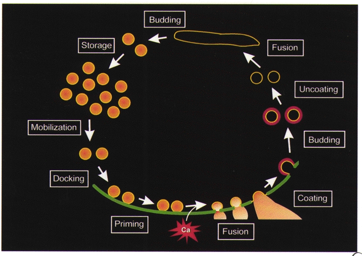
Neurotransmitter release relies on a series of synaptic vesicle trafficking reactions. We have determined the molecular basis of these reactions by microinjecting, into 'giant' nerve terminals of squid, probes that interfere with presynaptic proteins. These probes affect neurotransmitter release and disrupt nerve terminal structure. From the nature of these lesions, it is possible to deduce the roles of individual proteins in specific vesicle trafficking reactions. This approach has revealed the function of more than a dozen presynaptic proteins and we hypothesize that neurotransmitter release requires the coordinated action of perhaps 50-100 proteins.
Figures

A, measurements of presynaptic (Vpre) and postsynaptic (Vpost) responses before (control), during (pepE) and after (recovery) microinjection of a peptide from domain E of squid synapsin. B, injection of peptide from domain E of rat synapsin I enhances synaptic depression, measured as the decline in the rate of rise of postsynaptic responses (EPSP) evoked by a 50 Hz train of presynaptic action potentials. EPSP slope was normalized to values measured for the first response in each train. C, electron micrographs of presynaptic terminals injected with an inert control peptide (left) or a peptide from domain E of rat synapsin I (pepE; right). D, spatial distribution of synaptic vesicles measured in 50 nm shells surrounding active zones of terminals following injection of peptide from domain E of rat synapsin I or a control peptide. E, relative spatial distribution of synaptic vesicles in terminals injected with peptide from domain E of rat synapsin I, determined from the data shown in D by dividing values for domain E-injected terminals by values measured in control terminals. Modified from Hilfiker et al. (1998).

References
-
- Almers W. Synapses. How fast can you get? Nature. 1994;367:682–683. - PubMed
-
- Augustine GJ, Burns ME, DeBello WM, Pettit DL, Schweizer FE. Exocytosis: Proteins and perturbations. Annual Review of Pharmacology and Toxicology. 1996;36:659–701. - PubMed
-
- Augustine GJ, Charlton MP, Smith SJ. Calcium action in synaptic transmitter release. Annual Review of Neuroscience. 1987;10:633–693. - PubMed
Publication types
MeSH terms
Substances
Grants and funding
LinkOut - more resources
Full Text Sources