. 1999 Oct 1;520 Pt 1(Pt 1):281-92.
doi: 10.1111/j.1469-7793.1999.00281.x.

Modification of activity-dependent increases in cerebellar blood flow by extracellular potassium in anaesthetized rats

Affiliations

Modification of activity-dependent increases in cerebellar blood flow by extracellular potassium in anaesthetized rats

K Caesar et al. J Physiol. .

Abstract

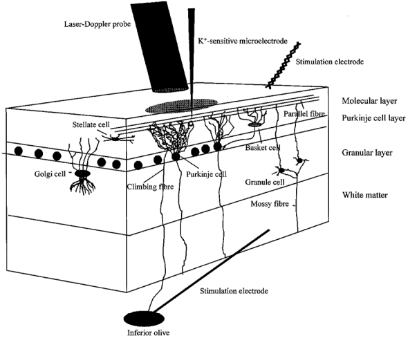
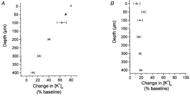
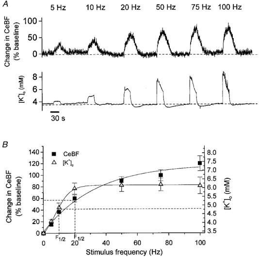
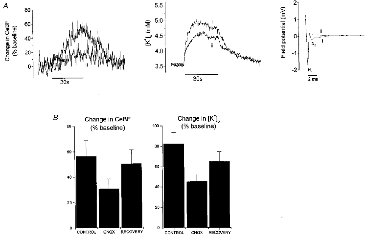
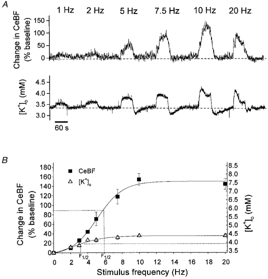
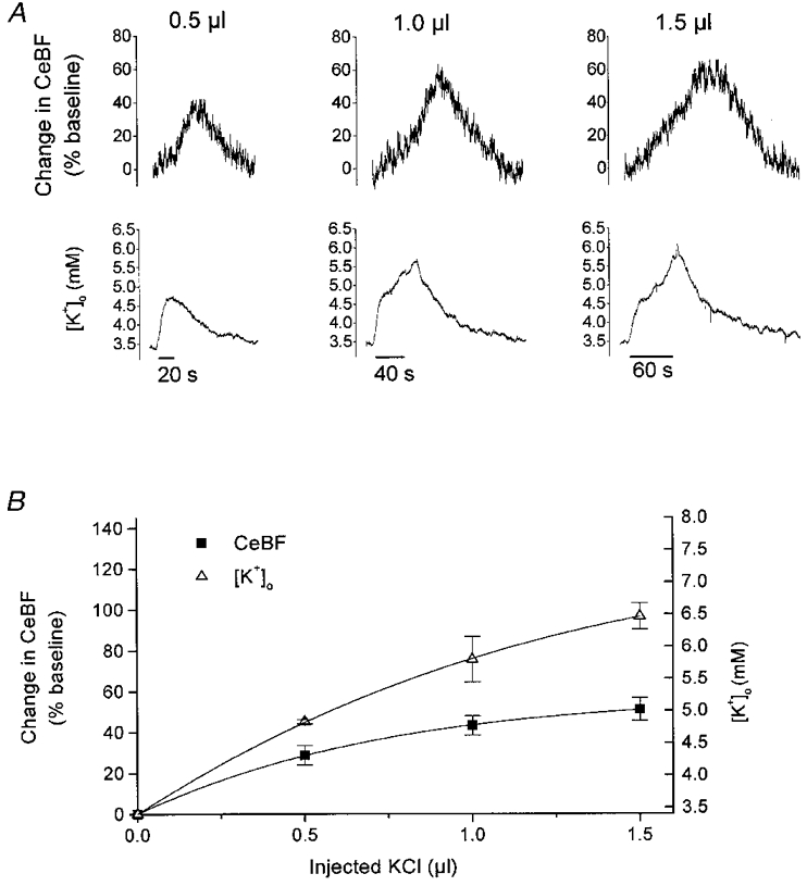
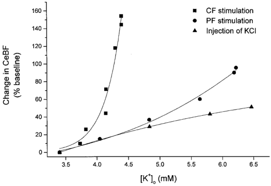
1. The hypothesis that potassium ions mediate activity-dependent increases of cerebral blood flow was examined in rat cerebellar cortex using ion-selective microelectrodes and laser-Doppler flowmetry. Increases of cerebellar blood flow (CeBF) and extracellular potassium concentration ([K+]o) were evoked by stimulation of parallel fibres and climbing fibres, and by microinjection of KCl into the cortex. 2. For parallel fibre stimulation, there was a maximal increase in [K+]o to 6.3 +/- 0.5 mM and in CeBF of 122 +/- 11 %. Climbing fibre stimulation gave a maximal increase in [K+]o to 4.4 +/- 0.2 mM and in CeBF of 157 +/- 20 %. This indicates different maxima for [K+]o and CeBF, dependent on the afferent system activated. 3. [K+]o and CeBF responses evoked by parallel or climbing fibre stimulation increased rapidly at the onset of stimulation, but exhibited different time courses during the remainder of the stimulation period and during return to baseline. 4. Microinjections of KCl into the cortex increased [K+]o to levels comparable to those evoked by parallel fibre stimulation. The corresponding CeBF increases were the same as, or smaller than, for parallel fibre stimulation, and much smaller than for climbing fibre stimulation. This suggests that mediators other than [K+]o are important for activity-dependent cerebral blood flow increases. 5. The present study showed that increased [K+]o is involved in CeBF regulation in the parallel fibre system, but is of limited importance for CeBF regulation in the climbing fibre system. The hypothesis that K+ is a major mediator of activity-dependent blood flow increases is probably not generally applicable to all brain regions and all types of neuronal stimulation.


Figures

Figure 1
Figure 1. Schematic drawing of the experimental set-up
A three-dimensional sagittal view of the rat cerebellar cortex, showing the neurones of interest and the position of the laser-Doppler probe, K+-sensitive microelectrode and stimulating electrodes. The superficial parallel fibres were stimulated by a bipolar electrode positioned on the cerebellar surface, while the climbing fibres were stimulated by a monopolar electrode lowered into the inferior olive. CeBF was recorded by a laser-Doppler flowmetry (LDF) probe located 0.5 mm above the pial surface, whereas changes in [K+]o were recorded with a K+-sensitive microelectrode lowered 50-100 μm into the cortex.
Figure 2
Figure 2. The laminar distribution of increases in [K+]o in response to parallel and climbing fibre activation
A, the laminar distribution of increases in [K+]o at cortical depths of 0, 50, 100, 200, 300 and 400 μm (n = 3) during parallel fibre stimulation at 20 Hz. B, the laminar distribution of increases in [K+]o evoked by activation of climbing fibres at 5 Hz (n = 3). Increases in [K+]o are given as percentage increases from baseline.
Figure 3
Figure 3. Frequency-dependent increases in CeBF and [K+]o in response to parallel fibre stimulation
A, typical example of CeBF and [K+]o increases evoked by parallel fibre stimulation at 5, 10, 20, 50, 75 and 100 Hz for 30 s. Upper panel shows original recordings of CeBF increases as a percentage of the baseline value. Lower panel shows simultaneous [K+]o traces (mm). Bar (at bottom) indicates the duration of stimulation. B, CeBF and [K+]o increases plotted versus stimulation frequency (n = 9). Increases in CeBF are shown on the ordinate to the left as percentage increases of baseline, while maximal values to which [K+]o increased are shown on the ordinate to the right. The smooth curves represent sigmodial curve fitting. The dashed lines show the frequencies at which the half-maximum increase occurred (F½).
Figure 4
Figure 4. Inhibition of postsynaptic AMPA receptors attenuates increases in CeBF and [K+]o in response to parallel fibre stimulation
A, original records of changes in CeBF and [K+]o, and the evoked field potential during parallel fibre stimulation at 20 Hz for 30 s. i, recordings during control conditions; ii, recordings after inhibition of the postsynaptic response with the glutamate (AMPA) receptor antagonist CNQX (100 μm, topical application for 30 min). Both the CeBF response (shown as percentage increase of baseline) and the [K+]o response (mm) were attenuated during the inhibition. Bars (below traces) indicate the duration of parallel fibre stimulation. The field potential is shown on the right (mV; mean of 100 sweeps). While the control field potential (i) consists of both a presynaptic component (N1) and a postsynaptic component (N2), the field potential measured after inhibition of the postsynaptic AMPA receptors (ii) had no postsynaptic component. B, CNQX significantly attenuated the increases in CeBF and [K+]o (P < 0.05, n = 6). Increases in CeBF and [K+]o are shown as percentage increases before (control) and after application of CNQX and after washout of CNQX (recovery).
Figure 5
Figure 5. Frequency-dependent increases in CeBF and [K+]o in response to climbing fibre stimulation
A, typical example of CeBF and [K+]o increases evoked by climbing fibre stimulation at 1, 2, 5, 7.5, 10 and 20 Hz for 60 s. Upper panel depicts recordings of CeBF increases as a percentage of the baseline value. Lower panel shows simultaneous [K+]o traces (mm). Bar (at bottom) indicates the duration of stimulation. B, CeBF and [K+]o increases plotted versus stimulation frequency (n = 5). Increases in CeBF are shown on the ordinate to the left as percentage increases of baseline and the maximal values to which [K+]o increased are shown on the ordinate to the right. The smooth curves represent sigmodial curve fitting. The dashed lines show the frequencies at which the half-maximum increase occurred (F½).
Figure 6
Figure 6. Increases in CeBF and [K+]o in response to microinjection of KCl into the cortex
A, typical example of CeBF and [K+]o increases in response to microinjection of 0.5, 1 and 1.5 μl KCl (20 mm in ACSF) into the cortex. Upper panel shows original recordings of the CeBF increases as a percentage of the baseline value. Lower panel shows simultaneous [K+]o traces (mm). Bars (at bottom) indicate the duration of the injection. B, amplitude of CeBF and [K+]o increases (n = 4) plotted versus KCl volume. Increases in CeBF are shown on the ordinate to the left as percentage increases of baseline, while maximal values to which [K+]o increased are shown on the ordinate to the right.
Figure 7
Figure 7. Increases in CeBF in response to climbing fibre and parallel fibre stimulation and microinjection versus corresponding increases in [K+]o
Scatter plot of the percentage increase in CeBF evoked by parallel fibre (PF) and climbing fibre (CF) activation and microinjection of KCl versus the simultaneously evoked [K+]o increase. The figure shows that the change in CeBF for a given value of [K+]o differs between the three types of stimulation.

References

    1. Akgoren N, Dalgaard P, Lauritzen M. Cerebral blood increases evoked by electrical stimulation of rat cerebellar cortex: relation to excitatory synaptic activity and nitric oxide synthesis. Brain Research. 1996;710:204–214. - PubMed
    1. Akgoren N, Fabricius M, Lauritzen M. Importance of nitric oxide for local increases of blood flow in rat cerebellar cortex during electrical stimulation. Proceedings of the National Academy of Sciences of the USA. 1994;91:5903–5907. - PMC - PubMed
    1. Akgoren N, Mathiesen C, Rubin I, Lauritzen M. Laminar analysis of activity-dependent increases of CBF in rat cerebellar cortex: dependence on synaptic strength. American Journal of Physiology. 1997;42:H1166–1176. - PubMed
    1. Berne RM, Winn HR, Rubio R. The local regulation of cerebral blood flow. Progress in Cardiovascular Diseases. 1981;24:243–260. - PubMed
    1. Betz E, Csornai M. Action and interaction of perivascular H+, K+ and Ca2+ on pial arteries. Pflügers Archiv. 1978;374:67–72. - PubMed

Publication types

MeSH terms

LinkOut - more resources