. 1999 Oct;121(2):647-56.
doi: 10.1104/pp.121.2.647.

Rates of sugar uptake by guard cell protoplasts of pisum sativum L. Related To the solute requirement for stomatal opening

Affiliations

Rates of sugar uptake by guard cell protoplasts of pisum sativum L. Related To the solute requirement for stomatal opening

G Ritte et al. Plant Physiol. 1999 Oct.

Abstract

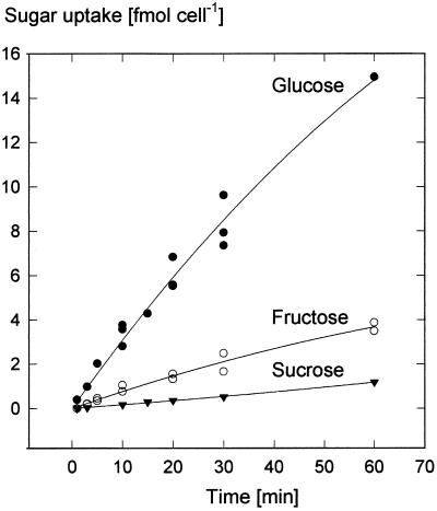
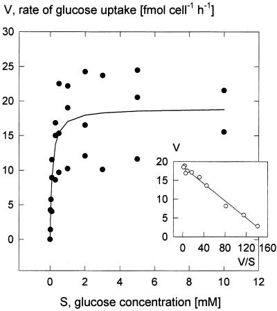
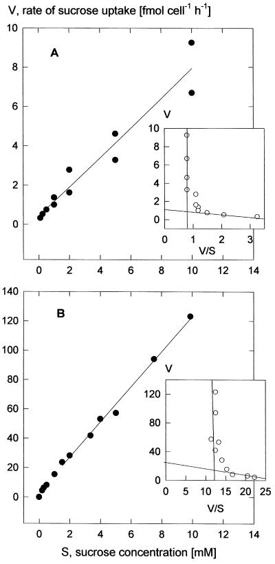
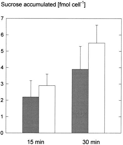
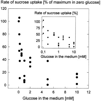
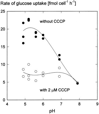
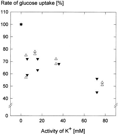
We wished to determine whether the capacity of the sugar uptake mechanisms of guard cells of the Argenteum mutant of pea (Pisum sativum L.) sufficed to support a concurrent stomatal opening movement. Sugar uptake by guard cell protoplasts was determined by silicone-oil-filtering centrifugation. The protoplasts took up [(14)C]glucose, [(14)C]fructose, and [(14)C]sucrose (Suc), apparently in symport with protons. Mannose, galactose, and fructose competed with Glc for transport by a presumed hexose carrier. The uptake of Glc saturated with a K(m) of 0.12 mM and a V(max) of 19 fmol cell(-1) h(-1). At external concentrations <1 mM, the uptake of Suc was slower than that of Glc. It exhibited a saturating component with a K(m) varying between 0.25 and 0.8 mM and a V(max) between 1 and 10 fmol cell(-1) h(-1), and at external concentrations >1 mM, a non-saturating component. At apoplastic sugar concentrations below 4 mM, sugar import was estimated to be mainly in the form of hexoses and too slow to support a simultaneous stomatal opening movement. If, however, during times of high photosynthesis and transpiration, the apoplastic Suc concentration rose and entered the range of non-saturating import, absorbed Suc could replace potassium malate as the osmoticum for the maintenance of stomatal opening.


Figures

Figure 1
Figure 1
Time courses of the uptake by guard cell protoplasts of [14C]Glc (n = 3, one datum only for uptake at 60 min), [14C]Fru (n = 2), and [14C]Suc (n = 3, the individual data are hidden behind the symbols). Uptake conditions were 0.5 mm sugar, pH 5.5, 20°C. Curves are second-order regressions. (In another series of experiments, Suc accumulation was higher than shown here; see Fig. 4.)
Figure 2
Figure 2
Concentration dependence of the rate of Glc uptake at pH 5.5 (15-min incubation time). Inset, Eadie-Hofstee plot of the means. Linear regression resulted in an apparent Km of 0.12 mm (ranging from 0.10–0.15 mm among three replications) and a mean Vmax of 19 fmol cell−1 h−1 (12–24 fmol cell−1 h−1).
Figure 3
Figure 3
Concentration dependencies of the rate of Suc uptake at pH 5.5 (30-min incubation time). A, Rates characteristic for the majority of the experiments; B, data from one set of experiments showing particularly high rates. The curves are second-order regressions. The Eadie-Hofstee: plots of the means (insets) bring out the saturating and linear concentration dependencies. Graphs parallel to the ordinates represent the linear components. Linear regression of the data for Suc concentrations <0.5 mm (A) gave apparent Km values between 0.18 and 0.28 mm, with a mean of 0.25 mm (three experiments), and in B for concentrations <1 mm apparent Km values between 0.4 and 1.2 mm, with a mean of 0.8 mm (three experiments).
Figure 4
Figure 4
Uptake of Suc in darkness and in the light. Guard cell protoplasts were incubated in 3 mm Suc at pH 5.5 and 20°C for 15 and 30 min in the dark (shaded bars) or in the light (white bars; 600 μmol m−2 s−1).
Figure 5
Figure 5
Rate of [14C]Suc uptake at pH 5.5 in the presence of increasing concentrations of unlabeled Glc (15-min incubation time) and a Suc concentration of 0.5 mm, pH 5.5. Rate of uptake in the absence of Glc was set at 100%. Inset, Semilogarithmic plot of the data.
Figure 6
Figure 6
Dependence of Glc uptake rate on the pH of the medium in the presence or absence of the uncoupler CCCP (2 μm). The Glc concentration in the incubation medium was 0.5 mm and incubation times were 5 min. CCCP in ethanol was added 1 min before the addition of [14C]Glc. In both treatments the ethanol concentration was 0.8%. The pH values of the MES-Tris buffers (33 mm) were determined with a pH electrode. Curves are third-order regressions.
Figure 7
Figure 7
Relative rates of Glc uptake as depending on the activity of K+ in the medium, counter ions were either Cl (▵) or SO42− (▾); establishment of the K+ diffusion potentials was ensured by the presence of 2.5 μm valinomycin. Osmolality was adjusted to 0.42 osmol by adding mannitol, and was determined with a vapor pressure osmometer (Wescor, Logan, Utah). The incubation media had a 0.5 mm Glc concentration; the pH was kept at 5.5 with 33 mm MES-Tris buffer. Incubation time was 10 min. During this time, changes in protoplast volume due to the absorption of K+ did not occur (according to determinations of the sorbitol-impermeable space; see “Materials and Methods”). Glc uptake rates in the absence of K+ were set to 100%.

Similar articles

Cited by

References

    1. Aked J, Hall JL. The uptake of glucose, fructose and sucrose into the lower epidermis of leaf discs of pea (Pisum sativum L. cv. Argenteum) New Phytol. 1993;123:271–276.
    1. Boorer KJ, Loo DDF, Frommer WB, Wright EM. Transport mechanism of the cloned potato H+-sucrose cotransporter StSUT1. J Biol Chem. 1996;271:25139–25144. - PubMed
    1. Borchert S, Grosse H, Heldt HW. Specific transport of inorganic phosphate, dihydroxyacetone phosphate and 3-phosphoglycerate into amyloplasts from pea roots. FEBS Lett. 1989;253:183–186.
    1. Borchert S, Harborth J, Schünemann D, Hoferichter P, Heldt HW. Studies of the enzymic capacities and transport properties of pea root plastids. Plant Physiol. 1993;101:303–312. - PMC - PubMed
    1. Buckhout TJ. Sucrose transport in isolated plasma-membrane vesicles from sugar beet (Beta vulgaris L.): evidence for an electrogenic sucrose-proton symport. Planta. 1989;178:393–399. - PubMed

LinkOut - more resources